. 2006 Nov-Dec;1759(11-12):562-72.
doi: 10.1016/j.bbaexp.2006.10.004. Epub 2006 Oct 21.

Dusty protein kinases: primary structure, gene evolution, tissue specific expression and unique features of the catalytic domain

Affiliations

Dusty protein kinases: primary structure, gene evolution, tissue specific expression and unique features of the catalytic domain

Jianbin Peng et al. Biochim Biophys Acta. 2006 Nov-Dec.

Abstract

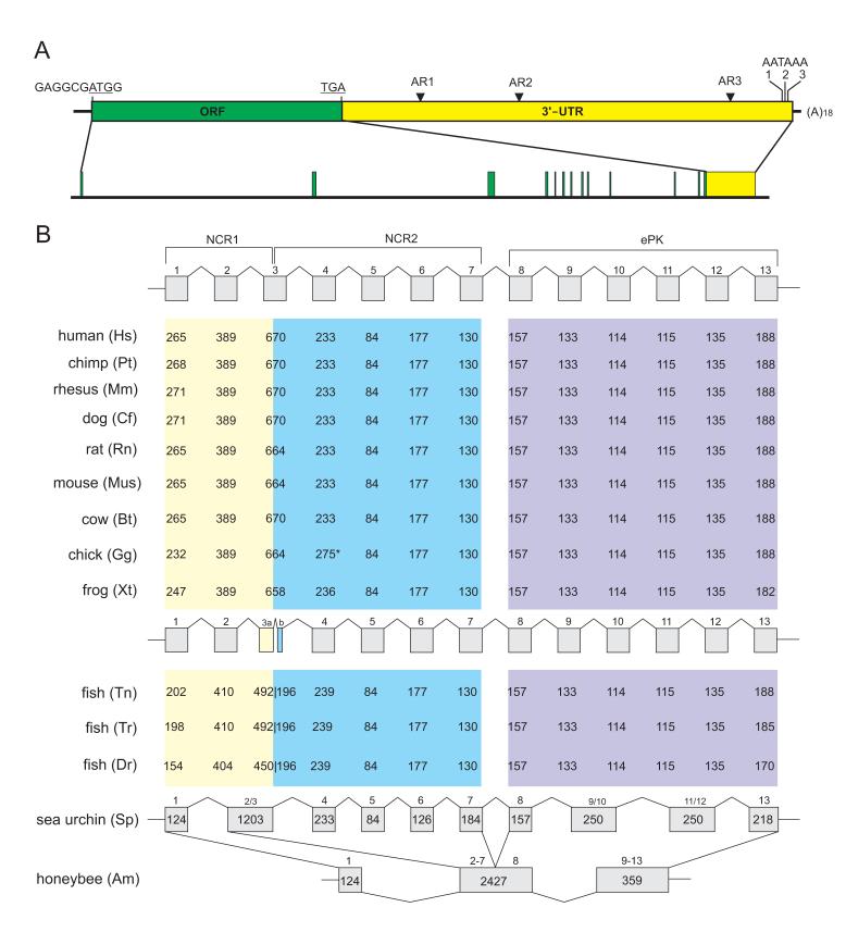
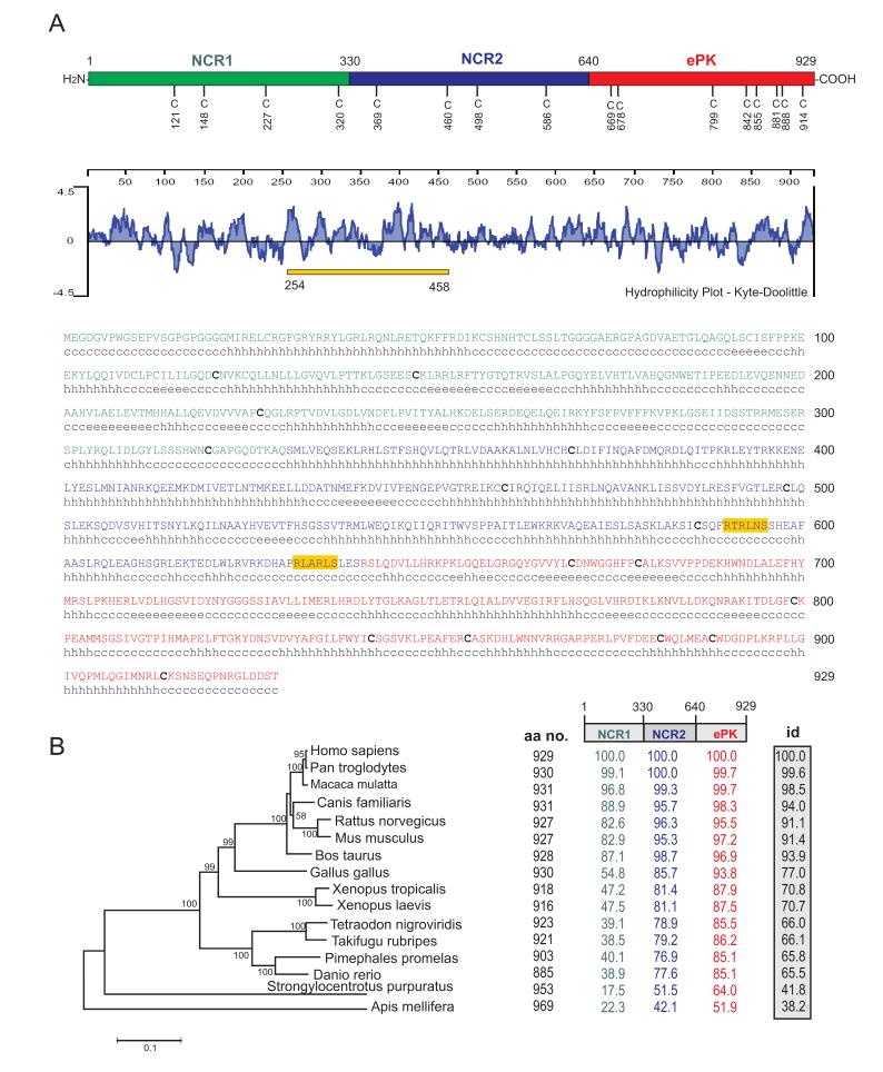
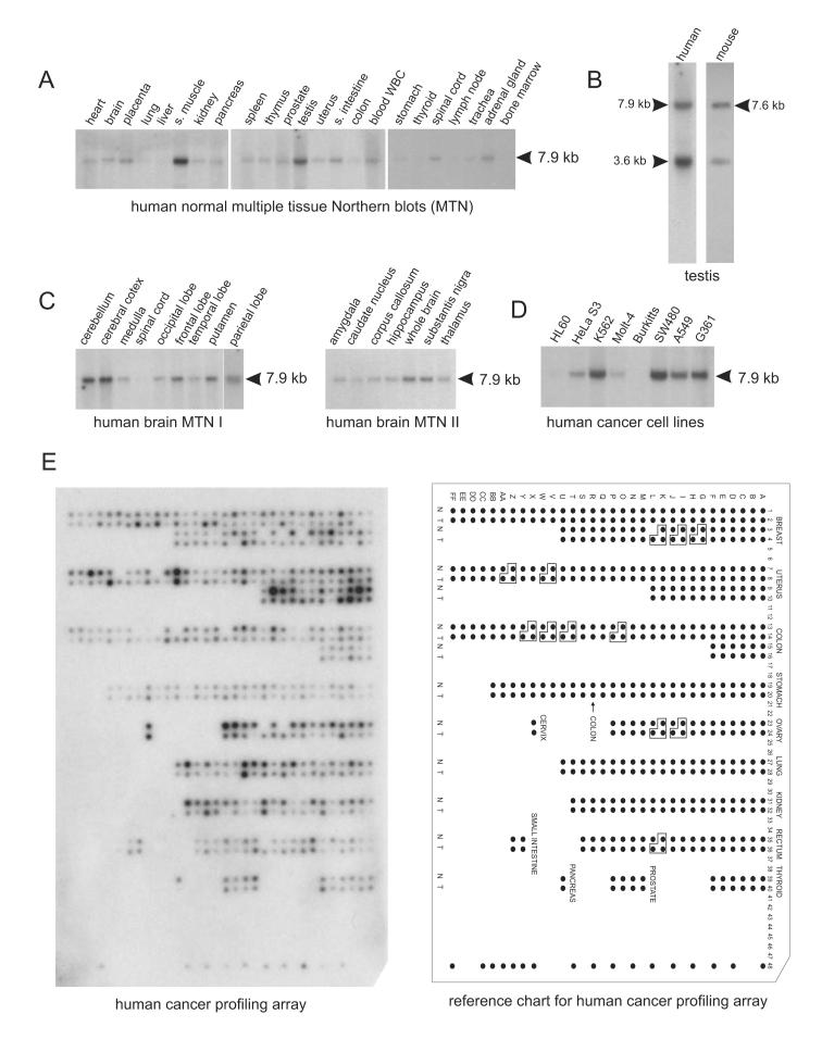
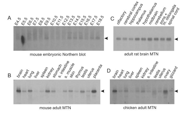
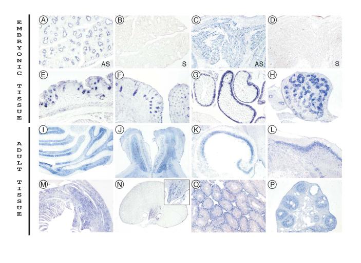
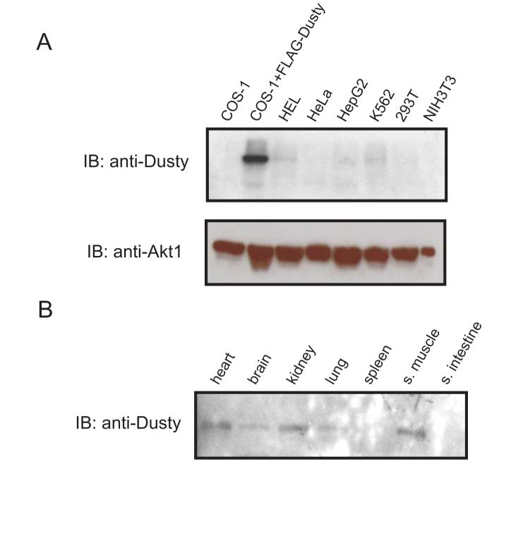
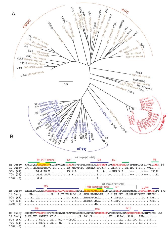
Ser/Thr- and Tyr-Protein kinases constitute a key switch underlying the dynamic nature and graded regulation of signal transduction and pathway activities in cellular organization. Here we describe the identification and characterization of Dusty, a single-copy gene that arose in metazoan evolution and encodes a putative dual Ser/Thr and Tyr protein kinase with unique structural features. Dusty is widely expressed in vertebrates, broadly distributed in the central nervous system, and deregulated in certain human cancers. Confocal imaging of transiently expressed human Dusty-GFP fusion proteins showed a cytoplasmic distribution. Dusty proteins from lower to higher species display an increasing degree of sequence conservation from the N-terminal non-catalytic domain to C-terminal catalytic domain. The non-catalytic region has eight conserved cysteine residues, multiple potential kinase-docking motifs and phosphorylation sites, whereas the catalytic domain is divergent and about equally distant of Ser/Thr and Tyr protein kinases. Homology analyses identified the essential catalytic residues, suggesting that Dusty homologues all possess the enzymatic activity of a protein kinase. Taken together, Dusty is a unique evolutionarily selected group of divergent protein kinases that may play important functional roles in the brain and other tissues of vertebrates.


Figures

Fig. 1
Fig. 1
Dusty gene structure and organization. (A) The mRNA structure and genomic organization of human Dusty as a model. The 7,898-base mRNA is divided into ORF, 3'UTR and poly(A) tail, where initiation and stop codons, Alu repeats (AR) and three polyadenylation signals (AATAAA) are shown. Division of mRNA into 13 exons is drawn to scale. (B) Exon-intron structure of Dusty in vertebrates. Exons (in bp) in correspondence to the protein sequence regions are denoted. Chicken exon 4 has an insertion (star-denoted) and fish exon 3 is split (3a and 3b). The organization of sea urchin Dusty is similar to that of vertebrates with fewer exon splits but is quite different from honeybee Dusty (bottom).
Fig. 2
Fig. 2
Dusty protein structure and evolution. (A) Human Dusty is shown as a model; it is divided in NCR1 (1-330), NCR2 (331-640) and ePK (641-929) domains, and contains 16 conserved Cys residues (whose positions are numbered) in vertebrates. The hydropathy plot, primary sequence and secondary structural elements (c, coiled-coil; e, extended strand; h, helix) are shown. The secondary structure shown for human Dusty is representative of other mammals, including chimp, rhesus monkey, cow, dog, rat and mouse. The bar denotes the peptide 254-458 for antibody production. Potential phosphorylation sites conserved in vertebrates are shaded (Akt/PKB: RTRLNS; and Pak: RLARLS). Conserved Cys residues are bolded. (B) The phylogram and pair-wise identity of Dusty proteins. Species names and bootstrap values are given (left). A scale bar denotes 0.2 substitutions per amino acid site. Shown also are the polypeptide size (total amino acids), domain identity (color-coded) and overall identity (shaded box) of each protein, as referred to human Dusty. Note the divergence of NCR1 among species.
Fig. 3
Fig. 3
Northern blot analysis of human Dusty. (A) Northern blots of 23 human tissues probed with Dusty-NB1 (exon 3-specific). The 7.9-kb band is denoted. (B) Northern blots of Dusty expressed in human (left) and mouse testis (right). Note a short mRNA form is seen in both species. (C) Northern blots of Dusty in human CNS regions. (D) Northern blots of Dusty in human cancer cell lines. In blots A to D (all from Clontech), each lane was loaded with 2 μg poly(A)+ RNA. Hybridization of actin probe was quite uniform and is not shown for brevity. (E) Human Cancer Profiling Array. The array (Clontech Cat#7841-1) was hybridized with the Dusty-CPA probe encoding the ePK domain (see supplementary table 2). N: normal tissue; and T: tumor tissue. The orientation grid for the array is shown at right. The identity and information of tumor tissue samples can be accessed on Clontech website or available from the authors upon request. Hybridization of the array with human ubiquitin cDNA was quite uniform and is not shown for brevity. Abbreviations: s. muscle, skeletal muscle; and s. intestine, small intestine.
Fig. 4
Fig. 4
Northern blot analysis of animal Dusty. (A) Northern blots of mouse embryonic development (E4.5 to E18.5) probed with Dusty-NB2. Note a basal expression in all stages and an elevation at E13.5 and onward. (B) Northern blots of mouse adult tissues. (C) Northern blots of rat adult brain probed with Dusty-NB3. Note Dusty is present in spinal cord. (D) Northern blots of adult chicken tissues probed with Dusty-NB4. All probes are exon 3 and species-specific. In blots A to D (all from SeeGene), each lane was loaded with 20 μg of total RNA and the loading was monitored with 28S and 18S rRNA bands. Hybridization of the blots with the actin or ubiquitin probe was quite uniform and is not shown for brevity.
Fig. 5
Fig. 5
Dusty RNA FISH in mouse embryonic and adult tissues. All the images were obtained with the P-I probe. A—H are for mouse embryonic tissues. A and B, E14.5 lung. C and D, E14.5 skeletal muscle. E —H, E18.5: E, skin; F, whisker; G, intestine; H, testis. A, C, E, F, G and H are obtained with the antisense (AS) probe, while B and D are controls of the sense (S) probe. I —P are for mouse adult tissues with the antisense probe. I—L are for the brain: I, cerebellum; J, olfactory; K, hippocampus; L, cerebral cortex. M, heart; N, kidney (inset: the high magnification of renal collecting tubules); O, testis; and P, ovary. Magnifications are 80x: A, B, C, D, E, H, O, P; 50x: F, N-inset; 40x: G, K, L, M; 20x: I, J; 8x: N.
Fig. 6
Fig. 6
Western blot analysis. The blots were probed with a polyclonal antibody against human Dusty254-458. (A) Endogenous Dusty detected in different cell lines with Dusty-specific antibody. The transiently expressed Flag-Dusty was used as positive control and Akt/PKBα as a loading control. (B) Mouse Dusty on multiple normal tissue blots detected with the human Dusty antibody.
Fig. 7
Fig. 7
Subcellular localization of GFP-tagged human Dusty. (A) The full-length GFP-Dusty and truncated forms are depicted, whose positions refer to human Dusty. (B) An image of pEGFP vector expressed in HEK293 cells as control was captured by confocal microscopy. (C) An image of full-length GFP-Dusty indicates the cytoplasmic location. (D) A representative image of GFP-Dusty truncated versions indicates the similar subcellular location, as shown above.
Fig. 8
Fig. 8
Phylogram and conservation pattern of Dusty ePK domains. (A) The ML optimal tree was reconstructed from 194 ePK (19 Dusty and 175 PSK+PTK) using JTT+4G+I model. The tree is not rooted. The ePK taxa of PSK, PTK and Dusty are marked with brown, blue and red, respectively. Except Dusty, other ePKs have known 3D structures. The ID and accession numbers of the total dataset are listed in supplemental table 4. The bar denotes 0.5 substitutions per amino acid site. The name and division of major protein kinase groups are denoted. (B) Functional motifs of ePK gleaned from the multiple sequence alignment between 19 Dusty and 175 PSK/PTK. The first row is human Dusty (1-256 equals to 651-906aa in Fig. 2A), the second row conserved residues among 19 Dusty (consensus), and third to fifth rows conserved residues among 194 ePK on 50, 70, and 100% levels. Specific motifs (M1 through M11) are marked in red. The ATP binding motif (M1) and catalytic core (M6a) are shaded in yellow. The conserved TP residues in the activation loop (M8) are denoted by stars. The α-helix and β-sheet are denoted with blue and green bars, respectively, while the remaining are loops.

References

    1. Kennelly PJ, Krebs EG. Consensus sequences as substrate specificity determinants for protein kinases and protein phosphatases. J. Biol. Chem. 1991;266:15555–15558. - PubMed
    1. Hunter T. Protein kinases and phosphatases: The Yin and Yang of protein phosphorylation and signaling. Cell. 1995;80:2 25–236. - PubMed
    1. Pawson T, Scott JD. Protein phosphorylation in signaling – 50 years and counting. Trends Biochem. Sci. 2005;30:286–290. - PubMed
    1. Hanks SK, Quinn A-M, Hunter T. The protein kinase family: conserved features and deduced phylogeny of the catalytic domains. Science. 1988;241:42–52. - PubMed
    1. Manning G, Whyte DB, Martinez R, Hunter T, Sudarsanam S. The protein kinase complement of the human genome. Science. 2002;298:1912–1934. - PubMed

Publication types

MeSH terms

Substances

Associated data