. 2007 Jan 18;229(3):186-93.
doi: 10.1016/j.tox.2006.10.014. Epub 2006 Oct 29.

Oxidative and nitrosative stress in trichloroethene-mediated autoimmune response

Affiliations

Oxidative and nitrosative stress in trichloroethene-mediated autoimmune response

Gangduo Wang et al. Toxicology. .

Abstract

Reactive oxygen and nitrogen species (RONS) are implicated in the pathogenesis of several autoimmune diseases. Also, increased lipid peroxidation and protein nitration are reported in systemic autoimmune diseases. Lipid peroxidation-derived aldehydes (LPDAs) such as malondialdehyde (MDA) and 4-hydroxynonenal (HNE) are highly reactive and bind proteins covalently, but their potential to elicit an autoimmune response and contribution to disease pathogenesis remain unclear. Similarly, nitration of protein could also contribute to disease pathogenesis. To assess the status of lipid peroxidation and/or RONS, autoimmune-prone female MRL+/+ mice (5-week old) were treated with trichloroethene (TCE), an environmental contaminant known to induce autoimmune response, for 48 weeks (0.5mg/ml via drinking water), and formation of antibodies to LPDA-protein adducts was followed in the sera of control and TCE-treated mice. TCE treatment led to greater formation of both anti-MDA- and -HNE-protein adduct antibodies and higher serum iNOS and nitrotyrosine levels. The increase in TCE-induced oxidative stress was associated with increases in anti-nuclear-, anti-ssDNA- and anti-dsDNA-antibodies. These findings suggest that TCE exposure not only leads to oxidative/nitrosative stress, but is also associated with induction/exacerbation of autoimmune response in MRL+/+ mice. Further interventional studies are needed to establish a causal role of RONS in TCE-mediated autoimmunity.


Figures

Fig. 1
Fig. 1
Anti-MDA- and anti-HNE-protein adduct antibodies in the serum of MRL+/+ mice treated with TCE. Anti-MDA-protein adduct antibodies (Fig. 1A) and anti-HNE-protein adduct antibodies (Fig. 1B) were determined by specific ELISAs. The results represent the means ± SD of six animals in each group. * P < 0.05 versus the vehicle-treated controls.
Fig. 1
Fig. 1
Anti-MDA- and anti-HNE-protein adduct antibodies in the serum of MRL+/+ mice treated with TCE. Anti-MDA-protein adduct antibodies (Fig. 1A) and anti-HNE-protein adduct antibodies (Fig. 1B) were determined by specific ELISAs. The results represent the means ± SD of six animals in each group. * P < 0.05 versus the vehicle-treated controls.
Fig. 2
Fig. 2
Nitrotyrosine levels in the serum of MRL +/+ mice treated with TCE. Values are means ± SD of six animals in each group. * p < 0.05 versus controls.
Fig. 3
Fig. 3
iNOS levels in the serum of MRL +/+ mice treated with TCE. Values are means ± SD of six animals in each group. * p < 0.05 versus controls.
Fig. 4
Fig. 4
Western blot analysis for iNOS expression in the livers of MRL +/+ mice. (A) iNOS expression in control mice (lanes 1–3) and TCE-treated mice (lanes 4–6). (B) Densitometric analysis of iNOS bands from control and TCE-treated mice. The results represent the means ± SD. * p < 0.05 versus controls.
Fig. 5
Fig. 5
Induction of autoantibodies in MRL+/+ mice treated with TCE. Serum levels of ANA (5A), anti-ssDNA antibodies (5B) and anti-dsDNA antibodies (5C). The results represent the means ± SD. * p < 0.05 versus controls.
Fig. 5
Fig. 5
Induction of autoantibodies in MRL+/+ mice treated with TCE. Serum levels of ANA (5A), anti-ssDNA antibodies (5B) and anti-dsDNA antibodies (5C). The results represent the means ± SD. * p < 0.05 versus controls.
Fig. 5
Fig. 5
Induction of autoantibodies in MRL+/+ mice treated with TCE. Serum levels of ANA (5A), anti-ssDNA antibodies (5B) and anti-dsDNA antibodies (5C). The results represent the means ± SD. * p < 0.05 versus controls.
Fig. 6
Fig. 6
The correlation of serum anti-MDA-protein adduct antibodies with ANA. The correlation was established by calculating correlation coefficients between anti-MDA-protein adduct antibodies and ANA.
Fig. 7
Fig. 7
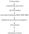
The anticipated sequence of events leading to autoimmune diseases following trichloroethene-induced oxidative stress.

References

    1. Ahmed SA, Penhale WJ, Talal N. Sex hormones, immune responses, and autoimmune diseases. Mechanisms of sex hormone action. Am J Pathol. 1985;121:531–551. - PMC - PubMed
    1. Ahsan H, Ali A, Ali R. Oxygen free radicals and systemic autoimmunity. Clin Exp Immunol. 2003;131:398–404. - PMC - PubMed
    1. Beckman JS, Koppenol WH. Nitric oxide, superoxide, and peroxynitrite: the good, the bad, and ugly. Am J Physiol. 1996;271:C1424–C1437. - PubMed
    1. Biemond P, Swaak AJ, Koster JF. Protective factors against oxygen free radicals and hydrogen peroxide in rheumatoid arthritis synovial fluid. Arthritis Rheum. 1984;27:760–765. - PubMed
    1. Channel SR, Latendresse JR, Kidney JK, Grabau JH, Lane JW, Steel-Goodwin L, Gothaus MC. A subchronic exposure to trichloroethylene causes lipid peroxidation and hepatocellular proliferation in male B6C3F1 mouse liver. Toxicol Sci. 1998;43:145–154. - PubMed

Publication types