Comparative Study
. 2007 May;129(1-2):35-45.
doi: 10.1016/j.pain.2006.09.033. Epub 2006 Nov 22.

Spinal NK-1 receptor expressing neurons mediate opioid-induced hyperalgesia and antinociceptive tolerance via activation of descending pathways

Affiliations
Comparative Study

Spinal NK-1 receptor expressing neurons mediate opioid-induced hyperalgesia and antinociceptive tolerance via activation of descending pathways

Louis P Vera-Portocarrero et al. Pain. 2007 May.

Abstract

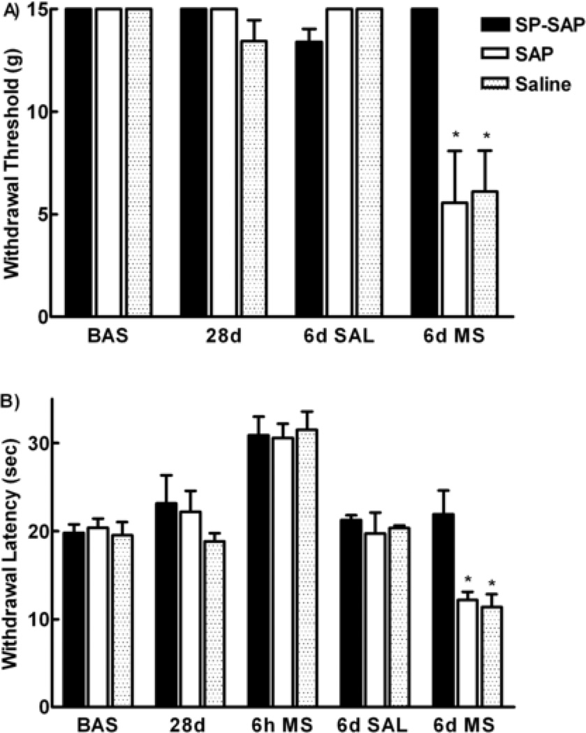
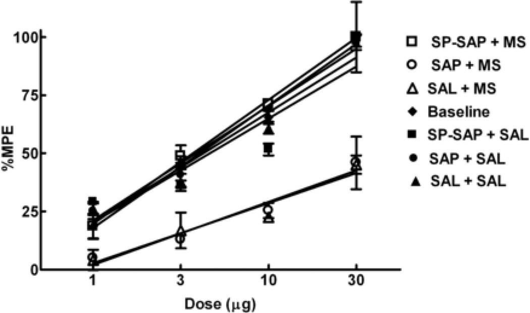
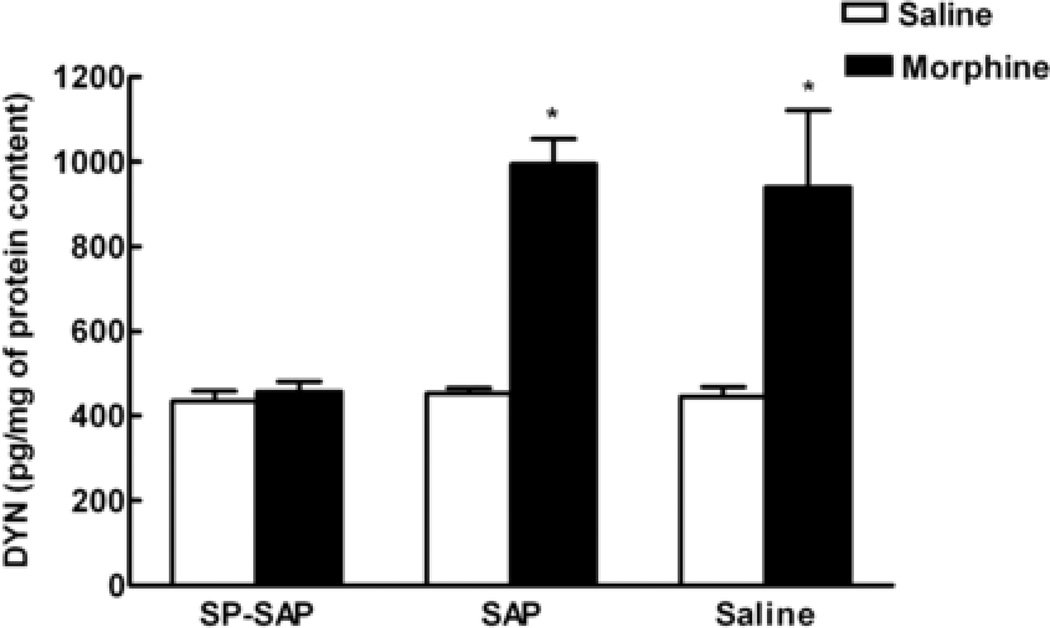
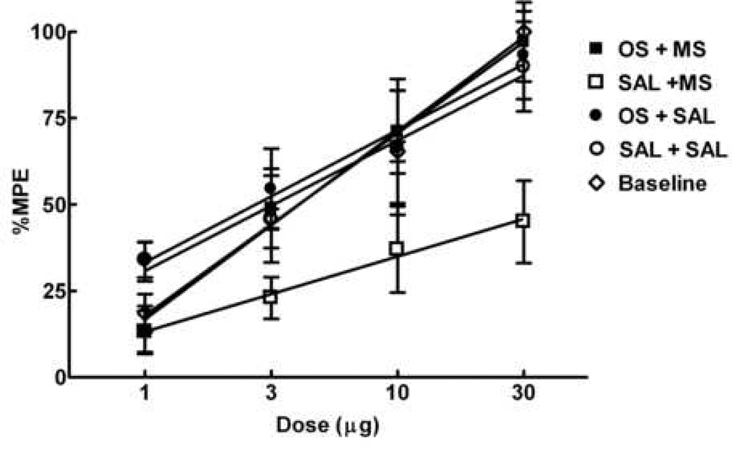
Opioids can induce hyperalgesia in humans and in animals. Mechanisms of opiate-induced hyperalgesia and possibly of spinal antinociceptive tolerance may be linked to pronociceptive adaptations occurring at multiple levels of the nervous system including activation of descending facilitatory influences from the brainstem, spinal neuroplasticity, and changes in primary afferent fibers. Here, the role of NK-1 receptor expressing cells in the spinal dorsal horn in morphine-induced hyperalgesia and spinal antinociceptive tolerance was assessed by ablating these cells with intrathecal injection of SP-saporin (SP-SAP). Ablation of NK-1 receptor expressing cells prevented (a) morphine-induced thermal and mechanical hypersensitivity, (b) increased touch-evoked spinal FOS expression, (c) upregulation of spinal dynorphin content and (d) the rightward displacement of the spinal morphine antinociceptive dose-response curve (i.e., tolerance). Morphine-induced hyperalgesia and antinociceptive tolerance were also blocked by spinal administration of ondansetron, a serotonergic receptor antagonist. Thus, NK-1 receptor expressing neurons play a critical role in sustained morphine-induced neuroplastic changes which underlie spinal excitability reflected as thermal and tactile hypersensitivity to peripheral stimuli, and to reduced antinociceptive actions of spinal morphine (i.e., antinociceptive tolerance). Ablation of these cells likely eliminates the ascending limb of a spinal-bulbospinal loop that engages descending facilitation and elicits subsequent spinal neuroplasticity. The data may provide a basis for understanding mechanisms of prolonged pain which can occur in the absence of tissue injury.


Figures

Figure 1
Figure 1
Mechanical and thermal hypersensitivity induced by sustained morphine exposure is prevented by SP-SAP. Male Sprague Dawley rats received a single i.th. injection of either SP-SAP, SAP or saline. Twenty–eight days later animals were implanted with morphine or saline minipumps. A) Mechanical threshold to stimulation with von Frey filaments was no different from baseline before implantation of the minipumps (28d). Six days after saline minipump implantation, the mechanical threshold did not change. Animals with morphine minipumps demonstrated a decrease in mechanical threshold (SAP and saline) (* denotes p< 0.05 compared to baseline and 28d values). In contrast animals with a single i.th. injection of SP-SAP had mechanical threshold similar to baseline. B) Paw withdraw latencies were no different from baseline before implantation of the minipumps (28d). Animals receiving morphine minipumps demonstrated analgesia represented by an increase in thermal latencies above baseline (6h ms). Six days after saline minipump implantation, the paw withdrawal latencies did not change. Animals with morphine minipumps demonstrated decreased paw withdrawal latencies in the SAP and saline groups (* denotes p < 0.05 compared to baseline and 28d values). In contrast, animals with a single i.th. SP-SAP injection had paw withdrawal latencies similar to baseline. There were 8 animals per group.
Figure 2
Figure 2
Antinociceptive tolerance to spinal morphine induced by sustained morphine exposure is prevented by SP-SAP. Male Sprague Dawley rats received i.th. injections of SP-SAP, SAP or saline and 28 days later either morphine (open symbols) or saline minipumps (closed symbols). Antinociceptive dose-response functions for i.th. morphine were generated before minipump implantation (Baseline) and 6 days after minipump implantation in the 52°C hot water tail-flick test. Each group of rats was tested with only one dose, 30 min after i.th. morphine injection. The dose-effect curve for i.th. morphine in groups with morphine minipumps and previous i.th. injections of SAP or saline was shifted significantly to the right of that for animals with saline minipumps (P<0.05). This dose-effect curve of animals with morphine minipumps and previous i.th. injection of SP SAP was not different from that of the animals with saline minipumps or baseline. There were 6 animals per dose.
Figure 3
Figure 3
The increase in dynorphin content in the spinal cord induced by sustained morphine exposure is prevented by SP-SAP. Male Sprague Dawley rats received single it.h injections of SP-SAP, SAP or saline and 28 days later implanted subcutaneously with morphine or saline (placebo) osmotic minipumps. At day 6 after minipump implantation, the spinal cords were removed, and the dorsal half of the lumbar cords were assayed for dynorphin content. The dorsal lumbar cords of rats with morphine minipumps and previous i.th. injections of SAP or saline showed significantly greater levels of dynorphin (* denotes P< 0.05 vs. saline groups) than tissues from rats with saline minipumps. The levels of spinal dynorphin from rats with saline minipumps were not different between the 3 groups (SP-SAP, SAP, saline). Morphine minipumps failed to significantly increase the levels of dynorphin in spinal tissues taken from animals with a previous single i.th. injection of SP-SAP. These levels were not different from those seen with saline minipumps. Each treatment group consisted of 8 animals.
Figure 4
Figure 4
Treatment with SP-SAP prevents the increase in touch-evoked FOS induced by sustained morphine exposure. Bar graph representing the number of FOS-positive cells in the spinal cord dorsal horn at the L5 levels counted in the side ipsilateral to tactile stimulation. Sprague Dawley rats received i.th. injections of SP-SAP, SAP or saline and 28 days later implanted with morphine or saline minipumps. The number of touch-evoked FOS positive cells in animals with morphine minipumps was increased compared with the number of FOS cells in animals with saline minipumps (* denotes P<0.05 vs. saline groups). This increase was only seen in animals with previous i.th. injection of SAP or saline. Animals with single i.th. injections of SP-SAP demonstrated similar numbers of touch-evoked FOS positive cells after morphine minipumps and saline minipumps. There were no differences in the number of touch-evoked FOS positive cells in the contralateral side to stimulation in any of the experimental groups.
Figure 5
Figure 5
Spinal administration of ondansetron reduces mechanical and thermal hypersensitivity induced by sustained morphine exposure. Male Sprague Dawley rats were implanted saline or morphine minipumps and 6 days later the effects of ondansetron on sustained morphine-induced hypersensitivity were studied. Rats with morphine minipumps demonstrated a decrease in mechanical (A) and thermal thresholds (B) at 6 days after minipump implantation. Injection of ondansetron (30 µg) it.h. attenuated the mechanical and thermal hypersensitivity 40 min after injection. It.h. injection of saline did not have any effects. (* denotes P<0.05 vs before it.h. injection). Animals with saline minipumps did not demonstrate changes in mechanical and thermal thresholds 6 days after implantation and after ondasentron injection.
Figure 6
Figure 6
Spinal administration of ondansetron reduces antinociceptive tolerance to spinal morphine induced by sustained morphine exposure. Male Sprague Dawley rats received either morphine (squares) or saline minipumps (circles). Antinociceptive dose-response functions for i.th. morphine were generated before minipump implantation (Baseline) and 6 days after minipump implantation in the 52°C water tail-flick test. Each group of rats was tested with only one dose, 30 min after i.th. morphine injection. The dose-effect curve for i.th. morphine in groups with morphine minipumps and previous i.th. injections of saline 10 min before morphine injection was shifted significantly to the right of that for animals with saline minipumps (P<0.05). The dose-effect curve of animals with morphine minipumps and previous i.th. injection of ondansetron (30 µg) was not different from that of the animals with saline minipumps or baseline. There were 6 animals per dose.

Similar articles

Cited by

References

    1. Abbadie C, Besson JM. C-fos expression in rat lumbar spinal cord following peripheral stimulation in adjuvant arthritic and normal rats. Brain Res. 1993;607:195–204. - PubMed
    1. Aley KO, Levine JD. Different mechanisms mediate development and expression of tolerance and dependence for peripheral mu-opioid antinociception in the rat. J Neurosci. 1997;17:8018–8023. - PMC - PubMed
    1. Ali NM. Hyperalgesic response in a patient receiving high concentrations of spinal morphine. Anesthesiology. 1986;65:449. - PubMed
    1. Angst MS, Clark JD. Opioid-induced hyperalgesia: A qualitative systematic review. Anesthesiology. 2006;3:570–587. - PubMed
    1. Arner S, Meyerson BA. Lack of analgesic effect of opioids on neuropathic and idiopathic forms of pain. Pain. 1986;33:11–23. - PubMed

Publication types

MeSH terms