. 2007 Feb;124(2):146-56.
doi: 10.1016/j.mod.2006.10.002. Epub 2006 Oct 11.

Impaired neural development caused by inducible expression of Axin in transgenic mice

Affiliations

Impaired neural development caused by inducible expression of Axin in transgenic mice

Hsiao-Man Ivy Yu et al. Mech Dev. 2007 Feb.

Abstract

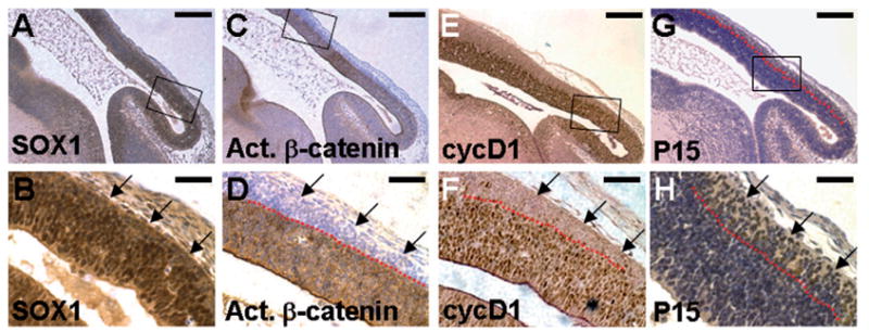
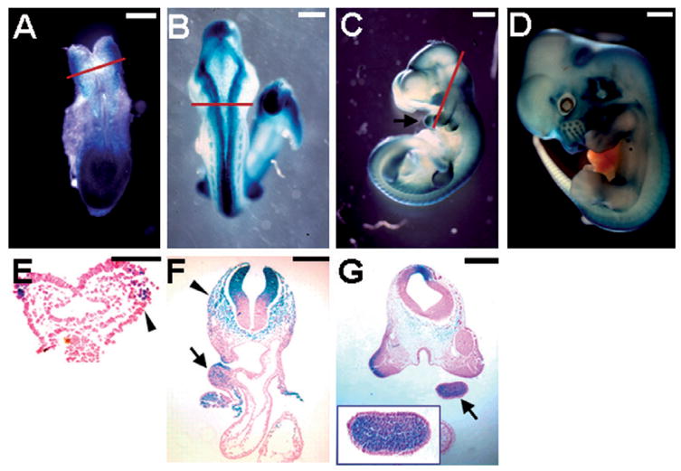
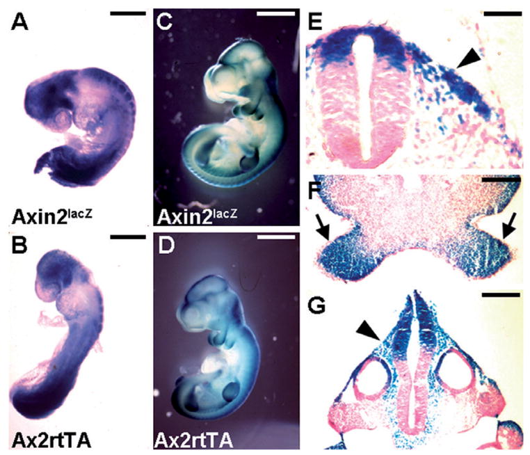
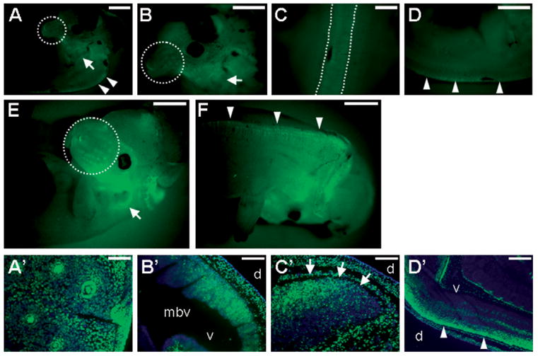
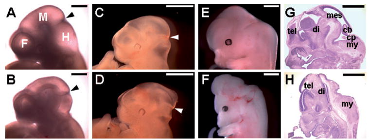
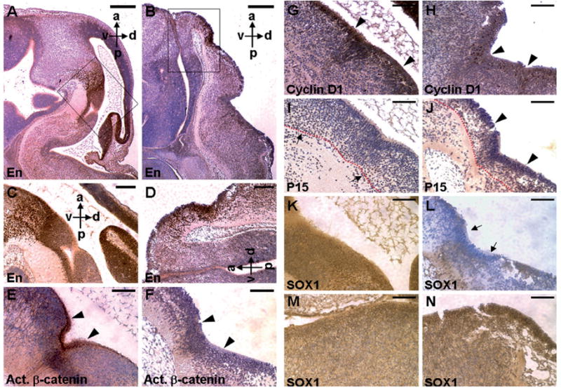
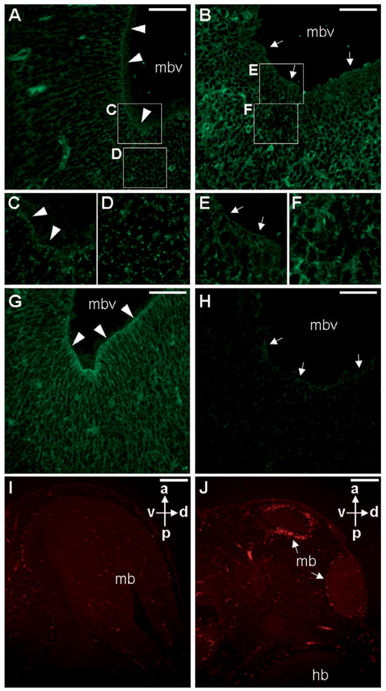
Ablations of the Axin family genes demonstrated that they modulate Wnt signaling in key processes of mammalian development. The ubiquitously expressed Axin1 plays an important role in formation of the embryonic neural axis, while Axin2 is essential for craniofacial skeletogenesis. Although Axin2 is also highly expressed during early neural development, including the neural tube and neural crest, it is not essential for these processes, apparently due to functional redundancy with Axin1. To further investigate the role of Wnt signaling during early neural development, and its potential regulation by Axins, we developed a mouse model for conditional gene activation in the Axin2-expressing domains. We show that gene expression can be successfully targeted to the Axin2-expressing cells in a spatially and temporally specific fashion. High levels of Axin in this domain induce a region-specific effect on the patterning of neural tube. In the mutant embryos, only the development of midbrain is severely impaired even though the transgene is expressed throughout the neural tube. Axin apparently regulates beta-catenin in coordinating cell cycle progression, cell adhesion and survival of neuroepithelial precursors during development of ventricles. Our data support the conclusion that the development of embryonic neural axis is highly sensitive to the level of Wnt signaling.


Figures

Fig. 1
Fig. 1
Wnt signaling maintains the mitotic activity of neuroepithelial precursors in developing ventricular zone. Sections of the E13.5 embryos were immunostained with α -SOX1 (A, B), α -ABC (C, D), α -cyclin D1 (E, F) or α -P15 (G, H) antibody (brown staining), and counterstained with hematoxylin (blue staining, A–D and G–H). Enlargements of the insets (A, C, E and G) are shown in B, D, F and H, respectively. Neuroepithelial precursors express SOX1 in the mid/hind brain regions (A, B). Immunohistochemical staining with an antibody that recognizes only the activated form of β -catenin (α -ABC antibody) reveals elevated levels of β -catenin in the neural precursors during brain development (C, D). Nuclear expression of cyclin D1 is evident in the developing ventricular zone (E, F). The red dashed lines represent the boundary of mitotic cells that express activated β -catenin/cyclin D1 and the postmitotic cells that do not (D.F). In contrast, P15 is present only in the post-mitotic cells (G, H). The arrows indicate SOX1-positive neuroepithelial cells, which exit the proliferating zone and express P15, but with no activation of β -catenin and cyclin D1. Scale bars, 0.2 mm (A, C, E and G); 50 μ m (B, D, F and H).
Fig. 2
Fig. 2
Expression of Axin2 in early neural development. The Axin2 expression pattern was visualized by β -gal staining in whole mounts or sections of the Axin2lacZ heterozygous embryos. At E8.5, Axin2 is expressed in the lateral margin of the neural fold (A) where neural crest cells arise at the boundary between the surface and neural ectoderm (E). By E9.5, strong Axin2 staining is then detected along the dorsal midline of the CNS (B). Axin2 is expressed in the dorsal neural tube, migrating neural crest cells (arrowheads) and branchial arches (arrows) (C, E, F and G). Inset shows an enlargement of the branchial arch of the E10.5 embryo (G). At E12.5, Axin2 is continuously present in the dorsal midline of CNS and many facial structures that are derivatives of CNC (D). First row, the Axin2lacZ knock-in embryos show that Axin2 expression. Second row, cross sections of the β -gal stained embryos. Red lines in A, B and C represent levels of the sections in E, F and G, respectively. Scale bars, 0.2 mm (A, F); 0.3 mm (B, G); 0.5 mm (C); 0.8 mm (D); 0.1 mm (E).
Fig. 3
Fig. 3
Axin2-rtTA mouse strain permits targeted gene expression in the Axin2-expressing domains. Embryos carrying Axin2-rtTA and TRE-lacZ transgenes enable conditional expression of the lacZ target gene in the Axin2-expressing cells. The transgenic expression was induced by Dox treatment of the pregnant mothers at E4.5/5.5. Embryos were recovered and analyzed by β -gal staining in whole mounts (B, D) and sections (E, F and G). The expression pattern of the lacZ target gene (B, D) is extremely similar to that of endogenous Axin2 gene detected using the Axin2lacZ knock-in allele at E9 (A) and E10.5 (C). The lacZ target gene is highly stimulated in the dorsal part of the neural tube, migrating neural crest cells (arrowheads) and branchial arches (arrows) upon Dox induction (E, F and G). Scale bars, 0.3 mm (A, B); 1 mm (C, D); 50 μ m (E); 0.2 mm (F, G).
Fig. 4
Fig. 4
Inducible expression of Axin1 in developing CNS and CNC. Embryos carrying the Axin2-rtTA, and either the TRE-Axin1-GFP or TRE-GFP transgene, display bi-cistronic expression of Axin1 and GFP (A–F) or expression of GFP (A′ –D′ ) in the Axin2-expressing cells. Transgene expression was induced by Dox treatment of the pregnant mothers beginning at E4.5/5.5. Embryos were analyzed by whole mount fluorescence microscopy for GFP at E12.5 (A–D) and E13.5 (E, F). The expression of the transgene was observed in the Axin2-expressing domain, including whisker hair follicles (dashed circle), external ears (arrows) and dorsal midline of CNS (dashed lines and arrowheads). Sections of the E13.5 embryos revealed expression of the target gene (GFP in green, counterstaining in blue) in whisker hair follicles (A′ ), midbrain (B′ ), hindbrain (C′ , arrows) and dorsal neural tube (D′ , arrowheads). mbv, midbrain ventricle; d, dorsal; v, ventral. Scale bars, 1 mm (A–F); 0.5 mm (C, D′ ); 0.1 mm (A′ ); 0.2 mm (B′ , C′ ).
Fig. 5
Fig. 5
High levels of Axin1 interfere with early brain development. Control embryos (A, C, E and G); Dox-induced transgenic embryos (B, D, F and H). Defects in the midbrain regions were noticeable in the E10.5 (A, B) and E11.5 (C, D) embryos with overexpression of Axin1 in the Axin2-expressing cells. Arrowheads indicate the mid/hindbrain boundary. At E13.5, severe brain abnormalities were detected (E, F). Histological analyses revealed that structures of the mescencephalon (mes), cerebellum (cb) and choroids plexus (cp) were completely missing (G, H). tel, telencephalon; di, diencephalon; my, myelencephalon. Control embryos (A, C, E and G); Dox-induced double transgenic embryos (B, D, F and H). Scale bars, 0.5 mm (A, B); 1 mm (C, D, E, F, G and H).
Fig. 6
Fig. 6
Development of the En-expressing domains is severely affected by Axin1, due to region-specific effects on Wnt mediated brain development. Sagittal sections of embryo at E13.5 were immunostained with specific antibodies (brown staining), and counterstained with hematoxylin (blue staining) as indicated. Immunohistochemical staining identifies the En-expressing domain in the control (A) and transgenic (B) midbrains. Enlargements of the insets (A, B) are shown in C and D, respectively. Panels E–L show the part of brain that is En-positive in adjacent sections (controls, E, G, I and K; transgenics, F, H, J and L). Overexpression of Axin1 affects β-catenin/cyclin D1 signaling in developing midbrain. Expression of the activated form of β-catenin (E, F) and cyclin D1 (G, H) is inhibited in the midbrain of transgenic embryos with elevated levels of Axin1. The arrowheads indicate that neuroepithelial progenitors, which are active mitotic cells with stimulated β-catenin and cyclin D1, are evident in the controls (E, G), but significantly reduced in the transgenics (F, H). P15, normally present in the post-mitotic cells (I, arrows), is detected in the proliferating ventricular zones of the mutants (J, arrowheads). The developing ventricular zone, indicated by the underlying red dashed line, is also reduced in the mutants. Immunohistochemical staining of SOX1 reveals specific inhibition of neural differentiation in the transgenic midbrains with high levels of Axin1. The SOX1-expressing neuroepithelial cells are detected in the control (K, M) and transgenic (L, N) midbrains (K, L) and hindbrains (M, N). High levels of Axin1 interfere with neural differentiation in the transgenic midbrain regions (arrows). a, anterior; p, posterior; d, dorsal; v, ventral. Scale bars, 500 μm (A, B); 400 μm (C, D); 200 μm (E–N).
Fig. 7
Fig. 7
Elevated levels of Axin1 affect formation of the actin microfilaments in the adherens junctions of developing ventricles, and survival of the neuroepithelial cells. Sections of the control (A, C, D and G) and transgenic (B, E, F and H) E13.5 embryos were analyzed by immunostaining of actin (A–F) or activated β-catenin (G, H), followed by confocal microscopy. Actin is localized to the adherens junctions of the control littermates (A, C) along the ventricle lumen of midbrain (arrowheads). In the transgenic embryos (B, E), distribution of the actin microfilaments is drastically reduced (arrows). However, the transgenic post-mitotic zone displays higher levels of actin filaments (D, F). Enlargements of the insets in A and B are shown in C–E and F. The activation of β-catenin is drastically reduced in the transgenic (arrows, H) compared to the control (arrowheads, G). TUNEL staining analyses revealed apoptotic cells in the control (I) and transgenic (J) E12.5 embryos. Increased numbers of apoptotic cells were detected in the transgenic midbrain ventricles (arrows). mb, midbrain; mbv, midbrain ventricle; hb, hindbrain; a, anterior; p, posterior; d, dorsal; v, ventral. Scale bars, 50 μm (A, B, G, H); 200 μm (I, J).

Similar articles

Cited by

References

    1. Behrens J, Jerchow BA, Wurtele M, Grimm J, Asbrand C, Wirtz R, Kuhl M, Wedlich D, Birchmeier W. Functional interaction of an axin homolog, conductin, with beta-catenin, APC, and GSK3beta. Science. 1998;280:596–599. - PubMed
    1. Behrens J, von Kries JP, Kuhl M, Bruhn L, Wedlich D, Grosschedl R, Birchmeier W. Functional interaction of beta-catenin with the transcription factor LEF-1. Nature. 1996;382:638–642. - PubMed
    1. Bhanot P, Brink M, Samos CH, Hsieh JC, Wang Y, Macke JP, Andrew D, Nathans J, Nusse R. A new member of the frizzled family from Drosophila functions as a Wingless receptor. Nature. 1996;382:225–330. - PubMed
    1. Bienz M. β-Catenin: a pivot between cell adhesion and Wnt signalling. Curr Biol. 2005;15:R64–R67. - PubMed
    1. Boutros M, Paricio N, Strutt DI, Mlodzik M. Dishevelled activates JNK and discriminates between JNK pathways in planar polarity and wingless signaling. Cell. 1998;94:109–118. - PubMed