. 2006 Dec 5;103(49):18597-602.
doi: 10.1073/pnas.0609061103. Epub 2006 Nov 21.

Dynamic assembly of end-joining complexes requires interaction between Ku70/80 and XRCC4

Affiliations

Dynamic assembly of end-joining complexes requires interaction between Ku70/80 and XRCC4

Pierre-Olivier Mari et al. Proc Natl Acad Sci U S A. .

Abstract

DNA double-strand break (DSB) repair by nonhomologous end joining (NHEJ) requires the assembly of several proteins on DNA ends. Although biochemical studies have elucidated several aspects of the NHEJ reaction mechanism, much less is known about NHEJ in living cells, mainly because of the inability to visualize NHEJ repair proteins at DNA damage. Here we provide evidence that a pulsed near IR laser can produce DSBs without any visible alterations in the nucleus, and we show that NHEJ proteins accumulate in the irradiated areas. The levels of DSBs and Ku accumulation diminished in time, showing that this approach allows us to study DNA repair kinetics in vivo. Remarkably, the Ku heterodimers on DNA ends were in dynamic equilibrium with Ku70/80 in solution, showing that NHEJ complex assembly is reversible. Accumulation of XRCC4/ligase IV on DSBs depended on the presence of Ku70/80, but not DNA-PK(CS). We detected a direct interaction between Ku70 and XRCC4 that could explain these requirements. Our results suggest that this assembly constitutes the core of the NHEJ reaction and that XRCC4 may serve as a flexible tether between Ku70/80 and ligase IV.


Conflict of interest statement

The authors declare no conflict of interest.

Figures
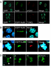
Fig. 1.
Fig. 1.
Accumulation of NHEJ proteins on laser-induced damage. (A) Accumulation of Ku80, EGFP-Ku80, γ-H2AX, and TUNEL staining in V79B cells after laser irradiation. The leftmost panels show the position of the laser-induced damage as dotted gray lines. (B) EGFP-Ku80 accumulation intensities correlate with DNA-dense heterochromatic regions. The chromatin density is revealed by the DNA dye DRAQ5 introduced after laser irradiation was performed. The image was taken 30 min after irradiation. (C) Accessibility of condensed chromosomes to DNA end binding of EGFP-Ku80. The image was taken 10 min after irradiation. (D) A representative cell division of EGFP-Ku80-expressing XR-V15B cells. The time in hours after irradiation is given in each frame. (Scale bars: 5 μm.)
Fig. 2.
Fig. 2.
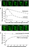
Ku70/80 interaction with DSBs in vivo. (A) Example of EGFP-Ku80 accumulation after laser irradiation of a disk inside the nucleus of XR-V15B cells expressing EGFP-Ku80. (B) Accumulation curve of EGFP-Ku80 on laser-induced DNA damage. For every cell, the pre-damage fluorescence level was set to 0 and the maximum was set to 1. The average and twice the SEM of a total of at least 10 cells is depicted in the graph. (Inset) Ku accumulation between 0 and 8 min in untreated cells shown in more detail. Filled circles represent untreated cells, and open triangles represent cells treated with Wortmannin. (C) Example of FRAP on a local EGFP-Ku80 accumulation. (D) FRAP on local damage curve for EGFP-Ku80. The data were normalized to the prebleach fluorescence level. The average and twice the SEM of 10 independent FRAP curves are depicted in the graph. The open circles represent the FRAP curve, and the filled circles show loss of fluorescence due to ongoing repair (from B). (Scale bars: 5 μm.)
Fig. 3.
Fig. 3.
Accumulation of NHEJ proteins on laser-induced damage. The immunofluorescent staining was done with antibodies to T2609 phosphorylated DNA-PKCS in HeLa cells or XRCC4 in V79B hamster cells. EGFP-ligase IV was directly imaged in primary human fibroblasts derived from a SCID patient with a ligase IV mutation that renders the endogenous protein unstable. (Scale bars: 5 μm.)
Fig. 4.
Fig. 4.
Interdependence of NHEJ protein assembly on DSBs. (A and B) Accumulation of Ku80 (green) in XRCC4-deficient XR-1 cells (A) and in DNA-PKCS mutant XR-C1 cells (B). (C and D) Accumulation of XRCC4 protein in XR-V15B cells expressing EGFP-Ku80 (C) and in DNA-PKCS-deficient XR-C1 cells (D). The arrowheads in C point to cells that do not express EGFP-Ku80. (Scale bars: 5 μm in A, B, and D and 20 μm in C.)
Fig. 5.
Fig. 5.
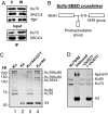
Direct interaction between Ku70 and XRCC4. (A) Immunoprecipitation from HeLa cell nuclear extracts using XRCC4 antisera. (Upper) The input material (10%). (Lower) A Western blot of the immunoprecipitated material. C, unirradiated cells; IR, cells irradiated with 45 Gy of γ-rays. (B) Schematic representation of the trifunctional cross-linker sulfo-SBED. (C) Cross-linked polypeptides after incubation of sulfo-SBED-labeled Ku70/80 with XRCC4, followed by photoactivation of the cross-linker. UV cross-linking was performed with only Ku70/80 (lane 1), only XRCC4 (lane 2), or Ku70/80 and XRCC4 (lanes 3 and 4). The products in lane 3 were treated with DTT to reverse the cross-link. The sizes of the molecular mass markers are depicted on the left, and the nature of the various products in the gel have been confirmed by mass spectrometry (MALDI-TOF). (D) Products that could be attached to the aryl azide group of sulfo-SBED that had been linked to Ku70/80. Mixtures contained Ku70/80, DNA-PKCS, ligase IV/XRCC4, and DNA. Lane 1 shows the Ku70/80 preparation alone, and lane 2 shows unbound proteins. Lane 4 shows the products that were precipitated by using streptavidin beads after activation of the aryl azide group and treatment with DTT, with lane 3 showing streptavidin-bound products without activation or DTT treatment.
Fig. 6.
Fig. 6.
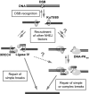
Working model for joining of various types of DNA DSBs. First, DSB recognition by Ku70/80 is fast and reversible. Subsequently, XRCC4/ligase IV can be attracted, forming a reversible complex. Alternatively, DNA-PKCS and XRCC4/ligase IV can both be recruited and form a more stable complex. We hypothesize that the joining reaction of simple DSBs can be accomplished via either of these two intermediates, whereas complex DSBs can be joined only via the right branch.

References

    1. van Gent DC, Hoeijmakers JH, Kanaar R. Nat Rev Genet. 2001;2:196–206. - PubMed
    1. Weterings E, van Gent DC. DNA Repair (Amst) 2004;3:1425–1435. - PubMed
    1. Smith GC, Jackson SP. Genes Dev. 1999;13:916–934. - PubMed
    1. Chan DW, Lees-Miller SP. J Biol Chem. 1996;271:8936–8941. - PubMed
    1. DeFazio LG, Stansel RM, Griffith JD, Chu G. EMBO J. 2002;21:3192–3200. - PMC - PubMed

Publication types