Global modulation of chromatin dynamics mediated by dephosphorylation of linker histone H1 is necessary for erythroid differentiation
- PMID: 17124174
- PMCID: PMC1656952
- DOI: 10.1073/pnas.0606478103
Global modulation of chromatin dynamics mediated by dephosphorylation of linker histone H1 is necessary for erythroid differentiation
Abstract
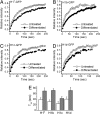
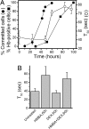
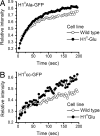
Differentiation of metazoan cells involves dramatic changes in gene expression patterns and proliferative capacity driven primarily by epigenetic mechanisms. Here we used in vivo photobleaching techniques and biochemical assays to investigate the contribution of alterations in chromatin dynamics to the differentiation of murine erythroleukemia (MEL) cells, a model system for erythroid development. As MEL cells differentiate the binding affinity of all linker histone variants increases, indicative of an overall decrease in chromatin flexibility. Changes in H1(0) binding properties depend on phosphorylation at one or more of three cyclin-dependent kinase sites. The presence of constructs mimicking constitutively phosphorylated H1 results in a significant inhibition in the acquisition of commitment to terminal cell division and the expression of erythroid-specific properties. These data indicate that the progressive loss of cdk activity associated with MEL cell differentiation leads to the accumulation of dephosphorylated linker histones and restricted chromatin flexibility and that these are necessary events in the progression of erythroid differentiation. We present additional data indicating that the presence of phosphorylated H1 has a dominant effect on the binding behavior of other linker histones and propose a model for the role of linker histone phosphorylation in which these modifications act within the context of assembled chromatin.
Conflict of interest statement
The authors declare no conflict of interest.
Figures


Similar articles
-
Role of H1 linker histones in mammalian development and stem cell differentiation.Biochim Biophys Acta. 2016 Mar;1859(3):496-509. doi: 10.1016/j.bbagrm.2015.12.002. Epub 2015 Dec 13. Biochim Biophys Acta. 2016. PMID: 26689747 Free PMC article. Review.
-
Remodeling somatic nuclei in Xenopus laevis egg extracts: molecular mechanisms for the selective release of histones H1 and H1(0) from chromatin and the acquisition of transcriptional competence.EMBO J. 1996 Nov 1;15(21):5897-906. EMBO J. 1996. PMID: 8918467 Free PMC article.
-
Site-specific regulation of histone H1 phosphorylation in pluripotent cell differentiation.Epigenetics Chromatin. 2017 May 22;10:29. doi: 10.1186/s13072-017-0135-3. eCollection 2017. Epigenetics Chromatin. 2017. PMID: 28539972 Free PMC article.
-
Plastrum testudinis induces γ-globin gene expression through epigenetic histone modifications within the γ-globin gene promoter via activation of the p38 MAPK signaling pathway.Int J Mol Med. 2013 Jun;31(6):1418-28. doi: 10.3892/ijmm.2013.1338. Epub 2013 Apr 8. Int J Mol Med. 2013. PMID: 23588991
-
Analyses of linker histone--chromatin interactions in situ.Biochem Cell Biol. 2006 Aug;84(4):427-36. doi: 10.1139/o06-071. Biochem Cell Biol. 2006. PMID: 16936816 Review.
Cited by
-
Photobleaching studies reveal that a single amino acid polymorphism is responsible for the differential binding affinities of linker histone subtypes H1.1 and H1.5.Biol Open. 2016 Feb 24;5(3):372-80. doi: 10.1242/bio.016733. Biol Open. 2016. PMID: 26912777 Free PMC article.
-
Developmentally regulated linker histone H1c promotes heterochromatin condensation and mediates structural integrity of rod photoreceptors in mouse retina.J Biol Chem. 2013 Jun 14;288(24):17895-907. doi: 10.1074/jbc.M113.452144. Epub 2013 May 3. J Biol Chem. 2013. PMID: 23645681 Free PMC article.
-
Overexpression of Rice Histone H1 Gene Reduces Tolerance to Cold and Heat Stress.Plants (Basel). 2023 Jun 22;12(13):2408. doi: 10.3390/plants12132408. Plants (Basel). 2023. PMID: 37446969 Free PMC article.
-
Role of H1 linker histones in mammalian development and stem cell differentiation.Biochim Biophys Acta. 2016 Mar;1859(3):496-509. doi: 10.1016/j.bbagrm.2015.12.002. Epub 2015 Dec 13. Biochim Biophys Acta. 2016. PMID: 26689747 Free PMC article. Review.
-
Analysis of the interplay between MeCP2 and histone H1 during in vitro differentiation of human ReNCell neural progenitor cells.Epigenetics. 2023 Dec;18(1):2276425. doi: 10.1080/15592294.2023.2276425. Epub 2023 Nov 17. Epigenetics. 2023. PMID: 37976174 Free PMC article.
References
-
- Eckfeldt CE, Mendenhall EM, Verfaillie CM. Nat Rev Mol Cell Biol. 2005;6:726–737. - PubMed
-
- Zhu L, Skoultchi AI. Curr Opin Genet Dev. 2001;11:91–97. - PubMed
-
- Hochedlinger K, Jaenisch R. Nature. 2006;441:1061–1067. - PubMed
-
- Becker PB. Nature. 2006;442:31–32. - PubMed
-
- Jenuwein T, Allis CD. Science. 2001;293:1074–1080. - PubMed
Publication types
MeSH terms
Substances
Grants and funding
LinkOut - more resources
Full Text Sources
Research Materials

