

A downloadable three-dimensional virtual model of the visible ear
- PMID: 17124433
- PMCID: PMC2655698
- DOI: 10.1159/000097369
A downloadable three-dimensional virtual model of the visible ear
Abstract
Purpose: To develop a three-dimensional (3-D) virtual model of a human temporal bone and surrounding structures.
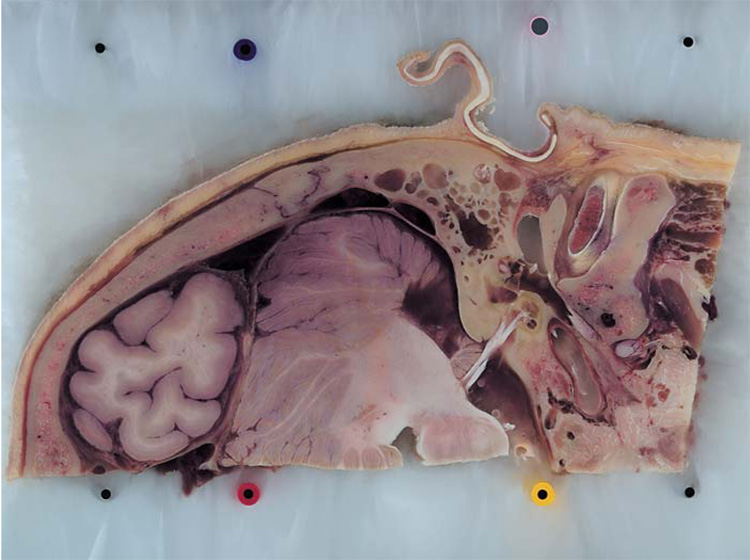
Methods: A fresh-frozen human temporal bone was serially sectioned and digital images of the surface of the tissue block were recorded (the 'Visible Ear'). The image stack was resampled at a final resolution of 50 x 50 x 50/100 micro m/voxel, registered in custom software and segmented in PhotoShop 7.0. The segmented image layers were imported into Amira 3.1 to generate smooth polygonal surface models.
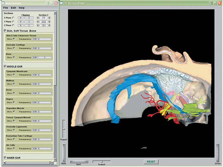
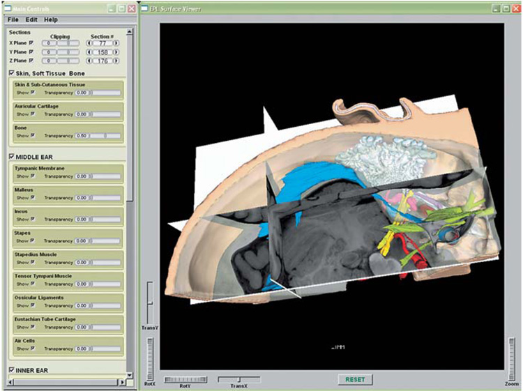
Results: The 3-D virtual model presents the structures of the middle, inner and outer ears in their surgically relevant surroundings. It is packaged within a cross-platform freeware, which allows for full rotation, visibility and transparency control, as well as the ability to slice the 3-D model open at any section. The appropriate raw image can be superimposed on the cleavage plane. The model can be downloaded at: (https://research.meei.harvard.edu/Otopathology/3dmodels/).
Figures
Similar articles
-
Three-dimensional virtual model of the human temporal bone: a stand-alone, downloadable teaching tool.Otol Neurotol. 2006 Jun;27(4):452-7. doi: 10.1097/01.mao.0000188353.97795.c5. Otol Neurotol. 2006. PMID: 16791035 Free PMC article.
-
The visible ear simulator: a public PC application for GPU-accelerated haptic 3D simulation of ear surgery based on the visible ear data.Otol Neurotol. 2009 Jun;30(4):484-7. doi: 10.1097/MAO.0b013e3181a5299b. Otol Neurotol. 2009. PMID: 19546800
-
Anatomy of the round window and hook region of the cochlea with implications for cochlear implantation and other endocochlear surgical procedures.Otol Neurotol. 2007 Aug;28(5):641-8. doi: 10.1097/mao.0b013e3180577949. Otol Neurotol. 2007. PMID: 17667773 Free PMC article.
-
Accessible and informative sectioned images, color-coded images, and surface models of the ear.Anat Rec (Hoboken). 2013 Aug;296(8):1180-6. doi: 10.1002/ar.22719. Epub 2013 May 27. Anat Rec (Hoboken). 2013. PMID: 23713007
-
The visible ear: a digital image library of the temporal bone.ORL J Otorhinolaryngol Relat Spec. 2002 Nov-Dec;64(6):378-81. doi: 10.1159/000066089. ORL J Otorhinolaryngol Relat Spec. 2002. PMID: 12499759
Cited by
-
The Usefulness of Reconstructed 3D Images in Surgical Planning for Cochlear Implantation in a Malformed Ear with an Abnormal Course of the Facial Nerve.Clin Exp Otorhinolaryngol. 2012 Apr;5 Suppl 1(Suppl 1):S48-52. doi: 10.3342/ceo.2012.5.S1.S48. Epub 2012 Apr 30. Clin Exp Otorhinolaryngol. 2012. PMID: 22701148 Free PMC article.
-
Using 3D Modeling Techniques to Enhance Teaching of Difficult Anatomical Concepts.Acad Radiol. 2016 Apr;23(4):507-16. doi: 10.1016/j.acra.2015.12.012. Epub 2016 Feb 17. Acad Radiol. 2016. PMID: 26897601 Free PMC article.
-
Virtual exploration and comparison of linear mastoid drilling trajectories with true-color volume rendering and the visible ear dataset.Stud Health Technol Inform. 2013;184:215-21. Stud Health Technol Inform. 2013. PMID: 23400159 Free PMC article.
-
Decentralized virtual reality mastoidectomy simulation training: a prospective, mixed-methods study.Eur Arch Otorhinolaryngol. 2019 Oct;276(10):2783-2789. doi: 10.1007/s00405-019-05572-9. Epub 2019 Jul 26. Eur Arch Otorhinolaryngol. 2019. PMID: 31350598
-
Three-dimensional histological specimen preparation for accurate imaging and spatial reconstruction of the middle and inner ear.Int J Comput Assist Radiol Surg. 2013 Jul;8(4):481-509. doi: 10.1007/s11548-013-0825-7. Epub 2013 Apr 30. Int J Comput Assist Radiol Surg. 2013. PMID: 23633112 Free PMC article.
References
-
- Takagi A, Sando I. Computer-aided three-dimensional reconstruction and measurement of the vestibular end-organs. Otolaryngol Head Neck Surg. 1988;98:195–202. - PubMed
-
- Takagi A, Sando I, Takahashi H. Computer-aided three-dimensional reconstruction and measurement of semicircular canals and their cristae in man. Acta Otolaryngol. 1989;107:362–365. - PubMed
-
- Nakashima S, Sando I, Tkahashi H, Fujita S. Computer-aided 3-D reconstruction and measurement of the facial canal and facial nerve. I. Cross-sectional area and diameter: preliminary report. Laryngoscope. 1993;103:1150–1156. - PubMed
-
- Harada T, Ishii S, Tayama N. Three-dimensional reconstruction of the temporal bone from histologic sections. Arch Otolaryngol Head Neck Surg. 1988;114:1139–1142. - PubMed
-
- Green JD, Jr, Marion MS, Erickson BJ, Robb RA, Hinojosa R. Three-dimensional reconstruction of the temporal bone. Laryngoscope. 1990;100:1–4. - PubMed
Publication types
MeSH terms
Grants and funding
LinkOut - more resources
Full Text Sources
