. 2006 Dec 13;25(24):5764-74.
doi: 10.1038/sj.emboj.7601436. Epub 2006 Nov 23.

A Cdt1-geminin complex licenses chromatin for DNA replication and prevents rereplication during S phase in Xenopus

Affiliations

A Cdt1-geminin complex licenses chromatin for DNA replication and prevents rereplication during S phase in Xenopus

Malik Lutzmann et al. EMBO J. .

Abstract

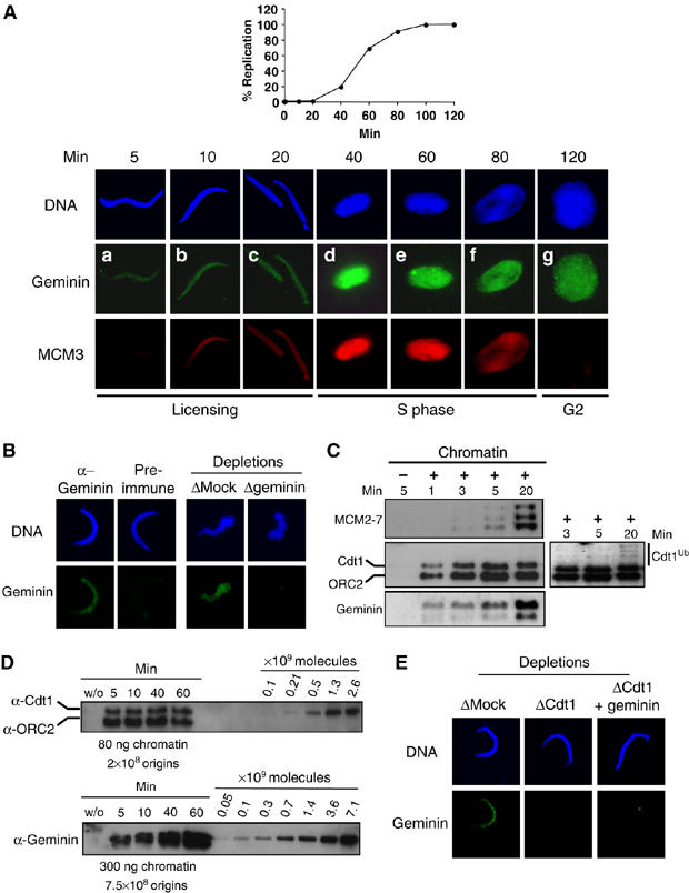
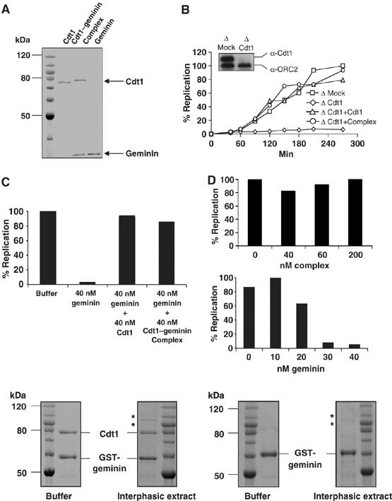
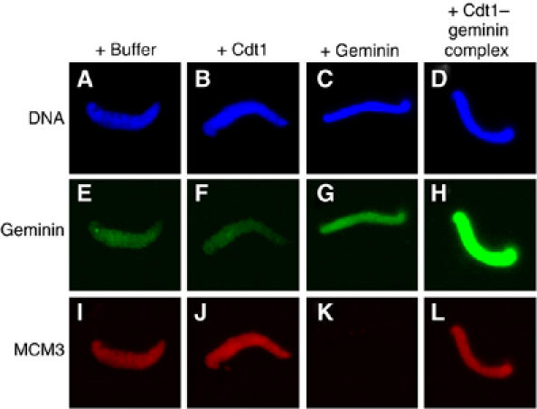
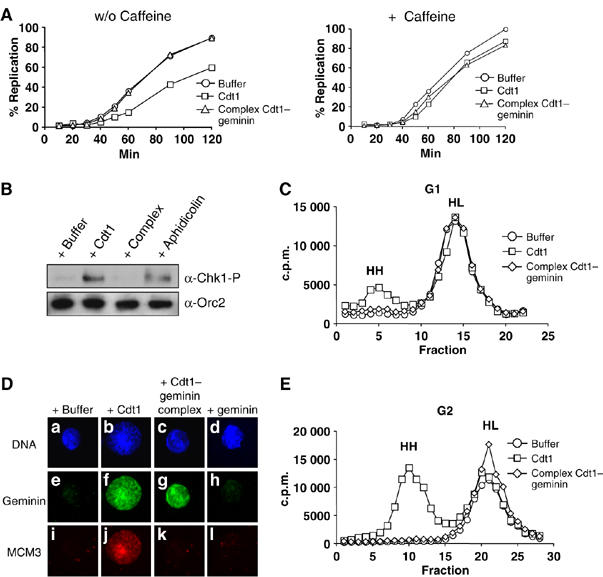
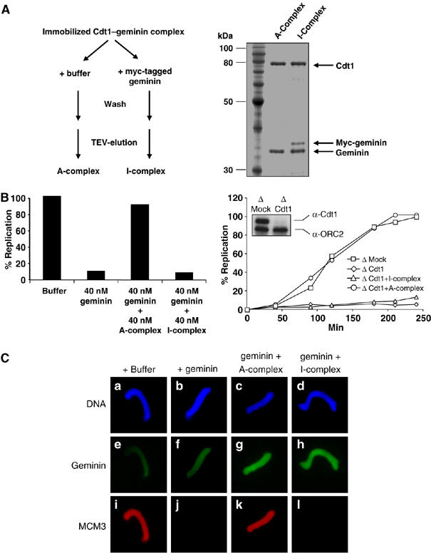
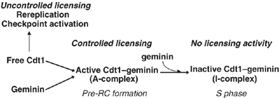
Initiation of DNA synthesis involves the loading of the MCM2-7 helicase onto chromatin by Cdt1 (origin licensing). Geminin is thought to prevent relicensing by binding and inhibiting Cdt1. Here we show, using Xenopus egg extracts, that geminin binding to Cdt1 is not sufficient to block its activity and that a Cdt1-geminin complex licenses chromatin, but prevents rereplication, working as a molecular switch at replication origins. We demonstrate that geminin is recruited to chromatin already during licensing, while bulk geminin is recruited at the onset of S phase. A recombinant Cdt1-geminin complex binds chromatin, interacts with the MCM2-7 complex and licenses chromatin once per cell cycle. Accordingly, while recombinant Cdt1 induces rereplication in G1 or G2 and activates an ATM/ATR-dependent checkpoint, the Cdt1-geminin complex does not. We further demonstrate that the stoichiometry of the Cdt1-geminin complex regulates its activity. Our results suggest a model in which the MCM2-7 helicase is loaded onto chromatin by a Cdt1-geminin complex, which is inactivated upon origin firing by binding additional geminin. This origin inactivation reaction does not occur if only free Cdt1 is present on chromatin.


Figures

Figure 1
Figure 1
A fraction of geminin assembles in a Cdt1-dependent manner during licensing onto chromatin. (A) Upper panel: kinetics of the replication reaction performed as described in Materials and methods and expressed as the percentage of replicated sperm DNA compared to the total input DNA. Lower panel: sperm chromatin incubated in egg extract for indicated times and further detergent extracted and processed for immunofluorescence. Geminin was visualized by FITC (green) and MCM3 by Texas red (red). DNA was stained with Hoechst and is shown in blue. (B) Left panel: immunofluorescence of chromatin purified as in (A) after 5 min incubation in egg extract and stained either with geminin antibody or with preimmune serum. Right panel: immunofluorescence of chromatin after 5 min incubation in egg extract that was either Mock- or geminin-depleted. (C) Western blot analysis of Cdt1, MCM2–7, geminin and ORC2 (as a loading control) on chromatin purified after the indicated time. Also shown is a Mock purification (first lane), without added sperm chromatin, to determine background staining by non-chromatin bound proteins. A longer exposure of the Cdt1 blot is also presented to show polyubiquitination of Cdt1 at the onset of S phase. (D) Western blot analysis of purified chromatin following the indicated times of incubation in egg extract (S-phase entry at 40 min). A dilution series of recombinant Cdt1 and geminin was loaded on the same gel. The amount of loaded chromatin is indicated, as well as the corresponding number of origins (Lemaitre et al, 2005). (E) Geminin signal on chromatin incubated 5 min in egg extract, either Mock-depleted, Cdt1-depleted, or Cdt1-depleted and complemented with geminin before addition of sperm chromatin.
Figure 2
Figure 2
The recombinant Cdt1–geminin complex is active in licensing DNA for replication. (A) Coomassie-stained SDS–PAGE of purified Cdt1, Cdt1–geminin complex and geminin. Also shown is a molecular weight marker. Protein concentrations were determined by comparison with BSA dilution series. (B) Kinetics of DNA replication in either Mock-depleted extract (squares) or in Cdt1-depleted extract supplemented by buffer (diamonds), 40 nM Cdt1 (triangles) or 40 nM Cdt1–geminin complex (circles). The insert shows a Western blot for Cdt1 and ORC2 (as loading control) of Mock- and Cdt1-depleted extract. (C) Percentage of replicated sperm chromatin after 100 min in egg extract supplemented by buffer, 40 nM geminin, 40 nM geminin plus 40 nM Cdt1, or 40 nM geminin plus 40 nM Cdt1–geminin complex. (D) Percentage of replicated sperm chromatin after 100 min in egg extract supplemented by buffer or by increasing amounts of Cdt1–geminin complex (upper panel) or by geminin (lower panel). (E) Incubation of Cdt1–geminin complex immobilized by GST-geminin to GSH-Sepharose (left panels) or GST-geminin alone immobilized in the same way (right panels) either in buffer or in interphasic egg extract for 40 min at room temperature (Coomassie stain). Additional bands visible after incubation in egg extract are proteins that bound nonspecifically to the GSH-Sepharose (marked by asterisks).
Figure 3
Figure 3
Geminin is massively recruited to chromatin without blocking MCM2–7 loading only if added in a complex with Cdt1. Immunofluorescence of geminin (FITC, green) and MCM3 (Texas red, red) on sperm chromatin (Hoechst, blue) after 5 min incubation in egg extract. As indicated, equal amounts of Cdt1, geminin or the Cdt1–geminin complex were added to the egg extract 10 min before the addition of sperm chromatin.
Figure 4
Figure 4
Free Cdt1, but not the Cdt1–geminin complex, leads to rereplication and checkpoint activation. (A) Left panel: kinetics of sperm chromatin replication supplemented with buffer (circles), 40 nM free Cdt1 (rectangles) or 40 nM Cdt1–geminin complex (triangles). Right panel: the same reactions were performed in the presence of 5 mM caffeine. (B) Western blot analysis of purified nuclei with a phospho-Chk1 antibody. Nuclei were purified after 100 min incubation of sperm chromatin in egg extract supplemented with either buffer, free Cdt1, Cdt1–geminin complex (all 40 nM), or Aphidicolin (50 μg/ml). A Western blot with ORC2 antibody as loading control is also shown. (C) Analysis of replicated DNA half-substituted with BrdUTP (‘heavy–light', HL) or double-substituted after rereplication (‘heavy–heavy', HH) on a CsCl gradient. Before sperm chromatin addition, reactions were supplemented either with buffer (circles), free Cdt1 (rectangles) or Cdt1–geminin complex (diamonds). (D) Immunofluorescence of G2 chromatin (120 min after sperm chromatin addition) and 20 min after addition of either buffer, free Cdt1, Cdt1–geminin complex, or free geminin. DNA is shown in blue stained with Hoechst, geminin in green (FITC) and MCM3 in red (Texas red). (E) Analysis of replicated DNA half-substituted with BrdUTP after one round of replication (‘heavy–light', HL) or double-substituted after rereplication (‘heavy–heavy', HH) on a CsCl gradient in reactions supplemented in G2 (120 min after sperm DNA was added) either with buffer (circles), free Cdt1 (rectangles) or Cdt1–geminin complex (diamonds).
Figure 5
Figure 5
The stoichiometry of the Cdt1–geminin complex determines its active and inactive state. (A) Left panel: diagram explaining the formation of the ‘A'- and ‘I'–Cdt1–geminin complex. Right panel: SDS–PAGE of the active A-complex and the inactive I-complex, which contains additional bound myc-tagged geminin. Also shown is a molecular weight marker. (B) Left panel: percentage of replicated sperm chromatin after 100 min in egg extract supplemented by buffer, 40 nM geminin, 40 nM geminin plus 40 nM A-complex, or 40 nM geminin plus 40 nM I-complex. Right panel: kinetics of DNA replication in either Mock-depleted extract (squares) or in Cdt1-depleted extract supplemented by buffer (diamonds), 40 nM A-complex (circles) or 40 nM I-complex (triangles). The insert shows a Western blot for Cdt1 and ORC2 (as loading control) of Mock- and Cdt1-depleted extract. (C) Immunofluorescence of sperm chromatin incubated for 20 min in egg extract complemented with buffer or equal amounts of either geminin alone, the A–Cdt1–geminin complex or I–Cdt1–geminin complex in addition to geminin. DNA is shown in blue (Hoechst), geminin in green (FITC) and MCM3 in red (Texas red).
Figure 6
Figure 6
Functional model of the Cdt1/geminin licensing switch on chromatin. Free Cdt1 can license DNA replication origins, but its activity is not controlled and leads to rereplication and S-phase checkpoint activation. In contrast, the Cdt1–geminin A-complex licenses origins in a precisely controlled manner, without rereplication or checkpoint activation. After nuclear import renders free geminin competent to interact with Cdt1 (Hodgson et al, 2002), the active A-complex incorporates additional geminin forming the I-complex blocked in further licensing activity. Degradation of Cdt1 and geminin during S phase and mitosis resets the system for another cycle.

Similar articles

Cited by

References

    1. Arias EE, Walter JC (2005) Replication-dependent destruction of Cdt1 limits DNA replication to a single round per cell cycle in Xenopus egg extracts. Genes Dev 19: 114–126 - PMC - PubMed
    1. Ballabeni A, Melixetian M, Zamponi R, Masiero L, Marinoni F, Helin K (2004) Human geminin promotes pre-RC formation and DNA replication by stabilizing CDT1 in mitosis. EMBO J 23: 3122–3132 - PMC - PubMed
    1. Bell SP, Dutta A (2002) DNA replication in eukaryotic cells. Annu Rev Biochem 71: 333–374 - PubMed
    1. Benjamin JM, Torke SJ, Demeler B, McGarry TJ (2004) Geminin has dimerization, Cdt1-binding, and destruction domains that are required for biological activity. J Biol Chem 279: 45957–45984 - PubMed
    1. Blow JJ, Laskey RA (1988) A role for the nuclear envelope in controlling DNA replication within the cell cycle. Nature 332: 546–548 - PubMed

Publication types