. 2006 Dec 13;25(24):5852-63.
doi: 10.1038/sj.emboj.7601425. Epub 2006 Nov 23.

Redundant functions of RIM1alpha and RIM2alpha in Ca(2+)-triggered neurotransmitter release

Affiliations

Redundant functions of RIM1alpha and RIM2alpha in Ca(2+)-triggered neurotransmitter release

Susanne Schoch et al. EMBO J. .

Abstract

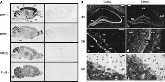
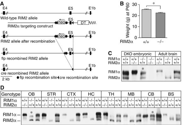
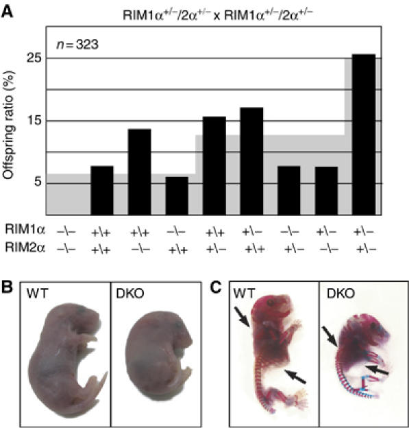
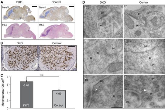
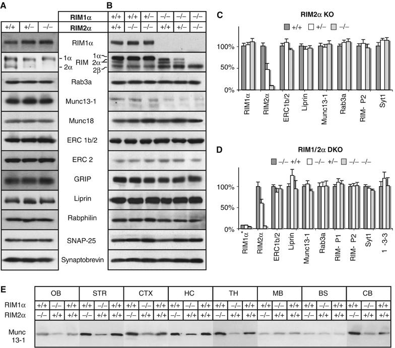
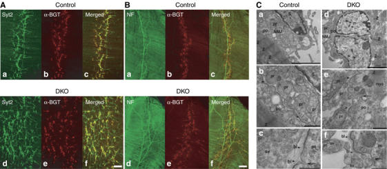
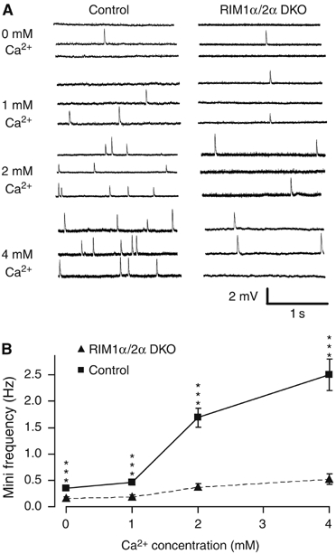
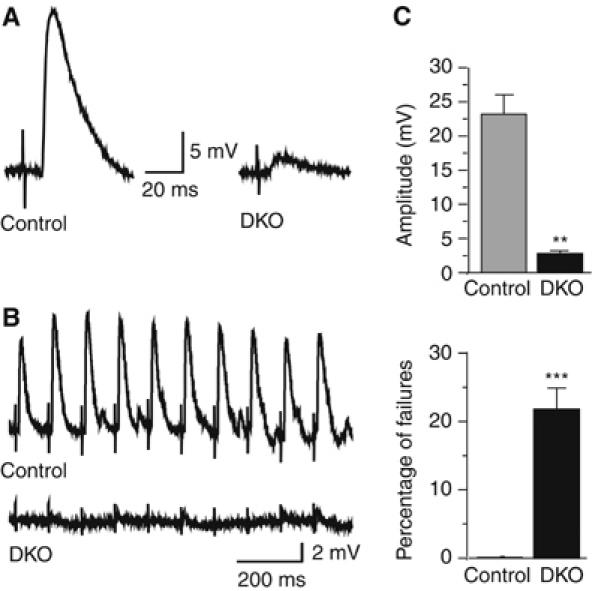
Alpha-RIMs (RIM1alpha and RIM2alpha) are multidomain active zone proteins of presynaptic terminals. Alpha-RIMs bind to Rab3 on synaptic vesicles and to Munc13 on the active zone via their N-terminal region, and interact with other synaptic proteins via their central and C-terminal regions. Although RIM1alpha has been well characterized, nothing is known about the function of RIM2alpha. We now show that RIM1alpha and RIM2alpha are expressed in overlapping but distinct patterns throughout the brain. To examine and compare their functions, we generated knockout mice lacking RIM2alpha, and crossed them with previously produced RIM1alpha knockout mice. We found that deletion of either RIM1alpha or RIM2alpha is not lethal, but ablation of both alpha-RIMs causes postnatal death. This lethality is not due to a loss of synapse structure or a developmental change, but to a defect in neurotransmitter release. Synapses without alpha-RIMs still contain active zones and release neurotransmitters, but are unable to mediate normal Ca(2+)-triggered release. Our data thus demonstrate that alpha-RIMs are not essential for synapse formation or synaptic exocytosis, but are required for normal Ca(2+)-triggering of exocytosis.


Figures

Figure 1
Figure 1
In situ hybridization of RIM mRNAs in the rat brain. (A) Film images showing the distribution of RIM mRNAs in the adult rat brain (CB, cerebellum; CX, cerebral cortex; DG, dentate gyrus; HC, hippocampus; MB, midbrain; OB, olfactory bulb; Pn, pontine nucleus; sTn, subthalamic nucleus; ST, striatum; TH, thalamus; MO medulla oblongata). (B) Dark-field images of emulsion-dipped sections from rat hippocampus, olfactory bulb, and cerebellum (AON, anterior olfactory nucleus; DG, dentate gyrus; EPL, external plexiform layer; GL, glomerular layer; GRL, granule cell layer; MCL, mitral cell layer; ML, molecular layer; PCL, Purkinje cell layer; scale bar B, a–d=100 μm, B, e–f 20 μm).
Figure 2
Figure 2
Generation of RIM2α KO mice. (A) Structures of the RIM2 wild-type gene (wild-type allele), of the targeting vector used for homologous recombination (targeting construct), and of the mutant RIM2 alleles after homologous recombination before and after further recombination of flp and cre recombinases. In the targeting vector, exon 5 is flanked by loxP sites (black triangles) and the neomycin resistance gene cassette (neo) that is flanked by flp recombination sites (black circles). A diphtheria toxin gene (DT) is included for negative selection. (B) Weights of male RIM2α KO and littermate control mice (N=36, *P<0.001). (C) Immunoblots of E18.5 embryonic and adult wild-type, RIM1α KO, RIM2α KO and embryonic DKO whole brain homogenates. (D) α-RIM immunoblots of proteins from different brain regions from adult RIM1α KO, RIM2α KO, and wild-type control mice; blots were probed with an antibody that recognizes both RIM1α and RIM2α (abbreviations of brain areas: OB, olfactory bulb; STR, striatum; CTX, cortex; HC, hippocampus; TH, thalamus; MB, midbrain, BS, brain stem; CB, cerebellum; *, bands of alternatively spliced RIM2α isoforms of higher molecular weight).
Figure 3
Figure 3
(A) Survival analysis of the offspring from matings of double heterozygous RIM1α/2α mutant mice. The black bars plot the observed frequency of the indicated genotypes as percentage of the total, whereas the gray background indicates the expected frequency based on Mendelian inheritance (N=323). (B, C) Images of E18.5 RIM1α/2α DKO mutant and control littermate mice overall morphology (B) and skeleton (C, bones are stained in blue and cartilage in pink; black arrows point to ribcage and cervical vertebrae).
Figure 4
Figure 4
Morphology of RIM1α/2α DKO brains and spinal cord. (A) H&E- and NeuN-stained sagittal section of brains from E18.5 mice of the indicated genotype (scale bar=2 mm). (B) NeuN-stained coronal sections of spinal cord from E18.5 mice of the indicated genotype (scale bar=100 μm). (C) Morphometric analysis of NeuN-immunopositive spinal motoneurons (**P<0.005). (D) Electron micrographs of synapses in the spinal cord. The arrows in (c, f) point to presynaptic dense projections in active zones. Abbreviations: pr, presynaptic; po, postsynaptic; sv, synaptic vesicles; pm, presynaptic plasma membrane. Scale bars, a=300 nm, d=370 nm, b=300 nm, e=240 nm, c=100 nm, f=130 nm.
Figure 5
Figure 5
Analysis of synaptic protein levels in brains from RIM2α KO mice and RIM1α/2α DKO embryos. Brain homogenates of the indicated genotypes were analyzed by immunoblotting using antibodies to the indicated proteins (A, B), the blots were quantified and all data normalized to control level (C, D, RIM1α level in (D) normalized to wild type). (A, C) RIM 2α-KO (adult), (B, D) RIM1α/RIM2α-DKO (E18.5). (E) Proteins from different brain areas of adult RIM1α KO, RIM2α KO, and wild-type mice were analyzed with an antibody against Munc13-1 (abbreviations of brain areas: OB, olfactory bulb; STR, striatum; CTX, cortex; HC, hippocampus; TH, thalamus; MB, midbrain; BS, brain stem; CB, cerebellum).
Figure 6
Figure 6
Morphology of diaphragm NMJs in RIM1α/2α DKO mice. (A, B) Confocal micrographs of E18.5 RIM1α/2α DKO mutant and control littermate whole-mount diaphragm muscles labeled with Texas Red-conjugated α-bungarotoxin (α-BGT) and antibodies to synaptotagmin-2 (Syt2) (A, a–d) or to neurofilament (NF) (B, a–d). (C) Electron micrographs of NMJs in RIM1α/2α DKO mice and control littermates demonstrate normal ultrastructure of NMJs in control and DKO mice. (a, d) Low-magnification electron micrographs of the NMJ demonstrating normal overall architecture of the NMJ in control and DKO mice. (b, c) and (e, f) Normal ultrastructural appearance of the NMJs at higher magnifications. Arrows point to postsynaptic invaginations that are not yet fully developed at E18 in both control and DKO mice. Arrowheads point to basal lamina material in the synaptic cleft of the NMJ. Abbreviations: NMJ, neuromuscular junction; S, Schwann cell; n, nucleus of a postsynaptic muscle fiber; Z, Z stripes in sarcomeres of postsynaptic muscle fibers; myo, myofibrils; pr, presynaptic; po, postsynaptic; bl, basal lamina of neuromuscular junction between the presynaptic terminals and the postsynaptic plasma membrane; sv, synaptic vesicles. Scale bars, A=100 μm, B=200 μm, C, a, d=3 μm, b, e=700 nm, c, f=400 nm.
Figure 7
Figure 7
Spontaneous miniature endplate potentials (mEPPs) at the diaphragm NMJ from control and RIM1α/2α DKO mice recorded in 40 mM KCl in 0–4 mM extracellular Ca2+. (A) Representative traces from muscles from control (left panel) and double-mutant mice (right panel). (B) Average mEPP frequencies as a function of extracellular Ca2+ concentration (data shown are means±s.e.m.; n (number of cells) for mutants/controls are 34/27 (0 μM Ca2+), 27/26 (1 μM Ca2+), 30/27 (2 μM Ca2+), and 26/22 (4 μM Ca2+), respectively, ***P<0.0005, Student's t-test).
Figure 8
Figure 8
Impaired evoked neurotransmitter release at the NMJ of RIM1α/2α DKO mice. (A, B) Representative traces of EPPs evoked at low frequency (A) or at 10 Hz (B) in muscles from control (left trace in (A), top trace in (B)) or RIM1α/2α DKO embryos (right trace in (A); bottom trace in (B)). (C) Decreased amplitudes (upper panel) and increased failure rates (lower panel) of evoked EPPs in α-RIM-deficient NMJs (for amplitudes (excludes failures), controls, n=3 embryos, 16 cells; DKO, n=4 embryos, 19 cells; for failure rates, controls=1 failure in 309 stimuli, N=3 mice, 25 cells; DKO=115 failures in 518 stimuli, N=4 mice, 39 cells, **P<0.001, ***P<0.0005, Student's t test).

Similar articles

Cited by

References

    1. Andrews-Zwilling YS, Kawabe H, Reim K, Varoqueaux F, Brose N (2006) Binding to Rab3A-interacting molecule RIM regulates the presynaptic recruitment of Munc13-1 and ubMunc13-2. J Biol Chem 281: 19720–19731 - PubMed
    1. Betz A, Thakur P, Junge HJ, Ashery U, Rhee JS, Scheuss V, Rosenmund C, Rettig J, Brose N (2001) Functional interaction of the active zone proteins Munc13-1 and RIM1 in synaptic vesicle priming. Neuron 30: 183–196 - PubMed
    1. Brandon EP, Lin W, D'Amour KA, Pizzo DP, Dominguez B, Sugiura Y, Thode S, Ko CP, Thal LJ, Gage FH, Lee KF (2003) Aberrant patterning of neuromuscular synapses in choline acetyltransferase-deficient mice. J Neurosci 23: 539–549 - PMC - PubMed
    1. Brose N, Hofmann K, Hata Y, Südhof TC (1995) Mammalian homologues of Caenorhabditis elegans unc-13 gene define novel family of C2-domain proteins. J Biol Chem 270: 25273–25280 - PubMed
    1. Calakos N, Schoch S, Südhof TC, Malenka RC (2004) Multiple roles for the active zone protein RIM1alpha in late stages of neurotransmitter release. Neuron 42: 889–896 - PubMed

Publication types

MeSH terms