. 2007 Jan;8(1):51-5.
doi: 10.1038/sj.embor.7400858. Epub 2006 Nov 24.

The supramolecular architecture of junctional microdomains in native lens membranes

Affiliations

The supramolecular architecture of junctional microdomains in native lens membranes

Nikolay Buzhynskyy et al. EMBO Rep. 2007 Jan.

Abstract

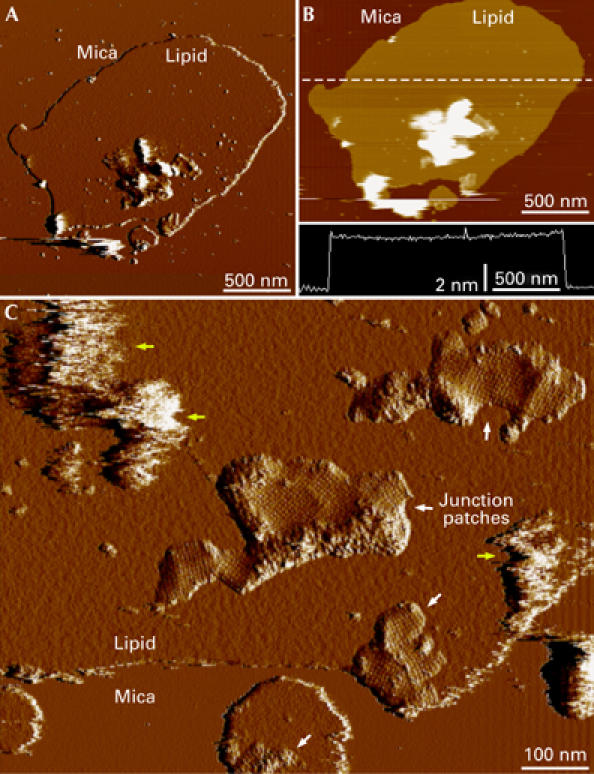
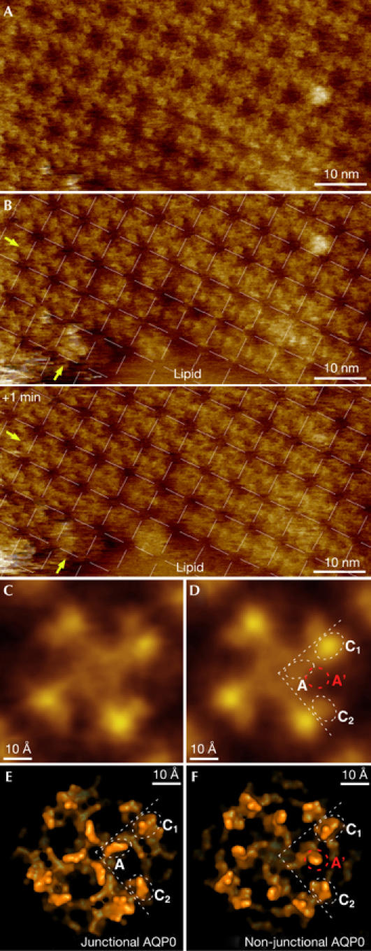
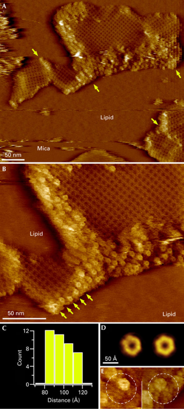
Gap junctions formed by connexons and thin junctions formed by lens-specific aquaporin 0 (AQP0) mediate the tight packing of fibre cells necessary for lens transparency. Gap junctions conduct water, ions and metabolites between cells, whereas junctional AQP0 seems to be involved in cell adhesion. High-resolution atomic force microscopy (AFM) showed the supramolecular organization of these proteins in native lens core membranes, in which AQP0 forms two-dimensional arrays that are surrounded by densely packed gap junction channels. These junctional microdomains simultaneously provide adhesion and communication between fibre cells. The AFM topographs also showed that the extracellular loops of AQP0 in junctional microdomains adopt a conformation that closely resembles the structure of junctional AQP0, in which the water pore is thought to be closed. Finally, time-lapse AFM imaging provided insights into AQP0 array formation. This first high-resolution view of a multicomponent eukaryotic membrane shows how membrane proteins self-assemble into functional microdomains.


Figures

Figure 1
Figure 1
Junctional microdomains within planar lipid bilayers. (A) Deflection and (B) height image of a native membrane patch isolated from the lens core. The height profile along the dashed line in (B) is shown in the lower panel and shows that the thickness of the bilayer is 46 Å. (C) Removal of vesicular structures that were attached to the membrane patches (yellow arrows) with the AFM tip showed the presence of corrugated patches that protruded from the lipid bilayer (white arrows). The patches contained square aquaporin 0 arrays.
Figure 2
Figure 2
Characterization of aquaporin 0 two-dimensional arrays. (A) High-resolution atomic force microscopy topograph of an aquaporin 0 (AQP0) array. (B) Time-lapse imaging of the border region of the same array with 1-min time interval (the AQP0 2D lattice is superimposed in both panels). AQP0 tetramers adjust to their lattice positions with time, as seen in the two edge rows of the array. Some tetramers move half a unit cell between the two images (indicated by arrows). (C) Unsymmetrized and (D) four-fold symmetrized AQP0 tetramer average topographs. (E) Surface representation of the junctional and (F) nonjunctional conformation of the extracellular AQP0 surface, which differ in the position of loop A.
Figure 3
Figure 3
High-resolution analysis of an aquaporin 0–connexon microdomain. (A) Topograph of aquaporin 0 (AQP0) arrays surrounded and separated (arrows) by densely packed connexon patches. (B) A higher magnification topograph that shows the characteristic flower shape of the connexons. The arrows indicate a row of connexons terminating an AQP0 array. (C) Histogram of distances between neighbouring connexons. (D) Unsymmetrized (left) and six-fold symmetrized connexon average topographies, calculated from 49 molecules. (E) AQP0–connexon pairs within unordered regions surrounding crystalline AQP0 arrays. In the left panel, the connexon is at the top right and AQP0 is at the bottom left of the circled area; in the right panel, the connexon is at the top left and AQP0 is at the bottom right of the circled area.

Similar articles

Cited by

References

    1. Agre P (2004) Nobel lecture. Aquaporin water channels. Biosci Rep 24: 127–163 - PubMed
    1. Alcala J, Lieska N, Maisel H (1975) Protein composition of bovine lens cortical fiber cell membranes. Exp Eye Res 21: 581–595 - PubMed
    1. Binnig G, Quate CF, Gerber C (1986) Atomic force microscope. Phys Rev Lett 56: 930–933 - PubMed
    1. Chandy G, Zampighi GA, Kreman M, Hall JE (1997) Comparison of the water transporting properties of MIP and AQP1. J Membr Biol 159: 29–39 - PubMed
    1. Donaldson P, Kistler J, Mathias RT (2001) Molecular solutions to mammalian lens transparency. News Physiol Sci 16: 118–123 - PubMed

Publication types

MeSH terms