. 2006 Dec;116(12):3127-38.
doi: 10.1172/JCI26620. Epub 2006 Nov 22.

Ca2+/calmodulin-dependent protein kinase II regulates cardiac Na+ channels

Affiliations

Ca2+/calmodulin-dependent protein kinase II regulates cardiac Na+ channels

Stefan Wagner et al. J Clin Invest. 2006 Dec.

Abstract

In heart failure (HF), Ca(2+)/calmodulin kinase II (CaMKII) expression is increased. Altered Na(+) channel gating is linked to and may promote ventricular tachyarrhythmias (VTs) in HF. Calmodulin regulates Na(+) channel gating, in part perhaps via CaMKII. We investigated effects of adenovirus-mediated (acute) and Tg (chronic) overexpression of cytosolic CaMKIIdelta(C) on Na(+) current (I(Na)) in rabbit and mouse ventricular myocytes, respectively (in whole-cell patch clamp). Both acute and chronic CaMKIIdelta(C) overexpression shifted voltage dependence of Na(+) channel availability by -6 mV (P < 0.05), and the shift was Ca(2+) dependent. CaMKII also enhanced intermediate inactivation and slowed recovery from inactivation (prevented by CaMKII inhibitors autocamtide 2-related inhibitory peptide [AIP] or KN93). CaMKIIdelta(C) markedly increased persistent (late) inward I(Na) and intracellular Na(+) concentration (as measured by the Na(+) indicator sodium-binding benzofuran isophthalate [SBFI]), which was prevented by CaMKII inhibition in the case of acute CaMKIIdelta(C) overexpression. CaMKII coimmunoprecipitates with and phosphorylates Na(+) channels. In vivo, transgenic CaMKIIdelta(C) overexpression prolonged QRS duration and repolarization (QT intervals), decreased effective refractory periods, and increased the propensity to develop VT. We conclude that CaMKII associates with and phosphorylates cardiac Na(+) channels. This alters I(Na) gating to reduce availability at high heart rate, while enhancing late I(Na) (which could prolong action potential duration). In mice, enhanced CaMKIIdelta(C) activity predisposed to VT. Thus, CaMKII-dependent regulation of Na(+) channel function may contribute to arrhythmogenesis in HF.


Figures
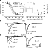
Figure 1
Figure 1. CaMKIIδc enhances steady-state inactivation of rabbit myocyte INa (10 mM [Na+]o).
(A) Mean INa availability (left) and INa during conditioning pulses (right; fit parameters in Table 1). In CaMKIIδc myocytes, availability was left-shifted versus β-gal (P < 0.05), and this was reversed by CaMKII inhibitors KN93 or AIP (P < 0.05). (B and C) Original INa traces during pre-pulses (right) and test pulse (left). INa amplitudes during pre-pulses were unaltered by CaMKIIδc (see Figure 2).
Figure 2
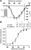
Figure 2. Em dependence of INa activation in rabbit myocytes (10 mM [Na+]o).
(A) Current-voltage relation (I-V) for CaMKIIδc versus β-gal myocytes. (B) INa activation curve, with relative conductance derived from maximal chord conductance and reversal potential (Erev) for each I-V, and peak INa/(Em – Erev). The resulting conductance was normalized to the maximal chord conductance (usually between –10 and 0 mV). CaMKIIδC had no effect on voltage for half-activation V1/2 or slope factor k (mV). Maximal INa was –4.3 ± 0.2 nA and –49.8 ± 2.4 pA/pF (Cm, 88.2 ± 5.7 pF) for CaMKIIδc versus –4.1 ± 0.5 nA and –42.8 ± 4.4 pA/pF (Cm, 103.8 ± 17.7 pF) for β-gal (P = NS).
Figure 3
Figure 3. CaMKIIδc increases IIM of INa in rabbit myocytes (10 mM [Na+]o).
(A) Increasing conditioning pulse duration (P1) reduced peak INa assessed with a second pulse (P2). CaMKIIδc increased the amount of accumulating IIM (versus β-gal; P < 0.05), an effect reversed by KN93 or AIP (P < 0.05). Mean data were fitted to a single exponential (see Table 1). (B) Original traces of peak INa (at P2) for different P1 durations.
Figure 4
Figure 4. CaMKIIδc slows INa recovery from inactivation in rabbit myocytes (10 mM [Na+]o).
(A) Increasing durations of recovery interval between conditioning pulse (P1, causing INa and inactivation) and test pulse (P2). CaMKIIδC significantly slowed recovery versus β-gal, an effect blocked upon CaMKII inhibition (KN93; P < 0.05). Elevation of [Ca2+]i to 500 nM in myocytes overexpressing CaMKIIδC further slowed recovery, and KN93 completely inhibited this effect. Mean data were fit to a single exponential (fit parameters in Table 1). (B) Original traces of INa for different recovery intervals.
Figure 5
Figure 5. CaMKIIδc slows fast decay of INa.
(A) Original traces show that CaMKIIδc slows fast INa inactivation in rabbit myocytes versus β-gal. INa decay (first 50 ms) was fit with a double exponential: y (t) = A1 exp (–t/τ1) + A2 exp (–t/τ2) + y0. Fits and τ1 and τ2 (ms) are shown in red (β-gal) and blue (CaMKIIδc). Deceleration of INa decay was significant in τ2 but not in τ1. KN93 could reverse the slowing of τ2 by CaMKIIδC. (B) In CaMKIIδC-Tg mice, both τ1 and τ2 were slowed compared with those in WT mice. Fits to the original traces and τ1 and τ2 values (ms) are shown in red (WT) and blue (CaMKIIδC-Tg). KN93 did not reverse these effects. Average peak INa was –10.2 ± 0.3 nA, –79.8 ± 2.7 pA/pF, Cm 132.7 ± 4 pF for rabbit and –14.2 ± 0.6 nA, –58.9 ± 2.9 pA/pF, Cm 255.3 ± 11 pF for mouse.
Figure 6
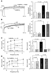
Figure 6. CaMKIIδc enhances late INa and increases [Na]i.
INa elicited at –20 mV (for 1,000 ms) was leak subtracted and normalized to peak INa. Current was integrated between 50 and 500 ms and normalized to the INa integral if no inactivation had occurred. (A) Original traces and mean data of normalized late INa. CaMKIIδC overexpression significantly increased late INa (reversed with KN93). (B) CaMKIIδC-Tg mice also showed late INa, but this was not reversible with KN93. Average peak INa was –10.2 ± 0.3 nA, –79.8 ± 2.7 pA/pF, Cm 132.7 ± 4 pF for rabbit and –14.3 ± 0.6 nA, –58.1 ± 2.8 pA/pF, Cm 259.7 ± 11.6 pF for mouse. (C) Mean [Na]i at different stimulation frequencies (left) and at 1 Hz (right) in rabbit myocytes. [Na]i was elevated in CaMKIIδc myocytes at all frequencies (P < 0.05) and reduced by KN93 (P < 0.05). (D) Mean data for CaMKIIδC-Tg mice also showing elevated [Na]i (versus WT; P < 0.05), but this was not reversed by KN93.
Figure 7
Figure 7. CaMKII-dependent association with and phosphorylation of Na+ channels.
(A and B) Equal amounts of CaMKII were IP from mouse hearts (A, Tg versus WT) and rabbit myocytes (B, CaMKIIδc versus nontransfected cells) and subjected to immunoblotting. CaMKII IP showed CaMKII association with Na+ channels in WT and Tg hearts (n = 3 and n = 3, respectively), while IP with polyclonal rabbit anti-Cav1.2a did not. Similar results were seen in rabbit myocytes (CaMKIIδC and nontransfected cells, n = 4 and n = 2, respectively). (C) Colocalization of CaMKII and Na+ channel in rabbit myocytes with immunocytological staining. (D and E) Autoradiograms measuring 32P incorporation into Na+ channels. Equal amounts of Na+ channels were immunoprecipitated from WT mouse hearts (n = 3) and directly phosphorylated with or without CaMKII, KN93 (50 μM), AIP (5 μM), or PKA/PKC inhibitor cocktail (PKA/C-I; 1 μM). (F) Endogenous CaMKII-dependent Na+ channel phosphorylation was activated in permeabilized rabbit myocytes by preincubating for 5 minutes in internal solutions of 500 nM [Ca2+] plus 2 μM CaM (versus 50 nM [Ca2+]). Sites not already phosphorylated were subsequently back-phosphorylated in Na+ channel immunoprecipitates (equal amounts) by exogenous preactivated CaMKII and ATP-γ-32P. The intensity of 32P is inversely related to the CaMKII-dependent phosphorylation during preincubation. (G and H) Western blots (anti-Nav Pan) show upregulation of Na+ channel expression in CaMKIIδC-Tg versus WT mice (relative to calsequestrin [CSQ]) but unaltered expression in CaMKIIδC rabbit myocytes (data pooled for MOI 10 and 100).
Figure 8
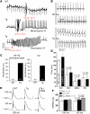
Figure 8. Arrhythmias in CaMKIIδC-Tg mice.
(A) Original ECG traces during programmed electrical stimulation in vivo. In WT mice, 2 consecutive premature beats (S2-S3, coupling interval 38 ms) did not induce arrhythmias (top). In contrast, in 2 different Tg mice, the same protocol induced monomorphic (middle) or polymorphic VT (bottom). (B) Representative ECG traces before and after isoproterenol (Iso) administration. P-waves (indicated by asterisks) were apparent during normal rhythms (WT with or without isoproterenol) but were absent in Tg mice after isoproterenol administration. This apparent AV dissociation indicates arrhythmia. (C) Frequency of arrhythmia induction was significant in Tg but not WT mice for both programmed electrical stimulation (left) and isoproterenol administration (right). (D) Resting ECG parameters (RR interval, corrected QT [QTc] interval, QRS duration, and PR interval). (E) Right-ventricular MAPs from hearts paced at basic cycle lengths (BCLs) of 100 (top) and 150 ms (bottom) for WT mouse hearts (left), Tg mouse hearts (middle), and Tg mouse hearts during infusion with KN93 (right). (F) Mean MAP durations at 90% repolarization (APD90). At slower heart rates (BCL, 150 ms), APD90 was significantly prolonged in Tg versus WT mice (but could not be reduced by KN93). At higher heart rates, there was no significant prolongation of APD90 in Tg versus WT.

References

    1. Bennett P., Yazawa K., Makita N., George A. Molecular mechanism for an inherited cardiac arrhythmia. Nature. 1995;376:683–685. - PubMed
    1. Wang D.W., Makita N., Kitabatake A., Balser J.R., George A.L. Enhanced Na+ channel intermediate inactivation in Brugada syndrome. . Circ. Res. 2000;87:E37–E43. - PubMed
    1. Bers, D.M. 2001. Excitation-contraction coupling and cardiac contractile force. Kluwer Academic Publishers. Dordrecht, The Netherlands. 427 pp.
    1. Tomaselli G.F., Zipes D.P. What causes sudden death in heart failure? Circ. Res. 2004;95:754–763. - PubMed
    1. Undrovinas A.I., Maltsev V.A., Sabbah H.N. Repolarization abnormalities in cardiomyocytes of dogs with chronic heart failure: role of sustained inward current. Cell. Mol. Life Sci. 1999;55:494–505. - PMC - PubMed

Publication types

MeSH terms

Substances