

Comparison of human uterine cervical electrical impedance measurements derived using two tetrapolar probes of different sizes
- PMID: 17125510
- PMCID: PMC1684260
- DOI: 10.1186/1475-925X-5-62
Comparison of human uterine cervical electrical impedance measurements derived using two tetrapolar probes of different sizes
Abstract
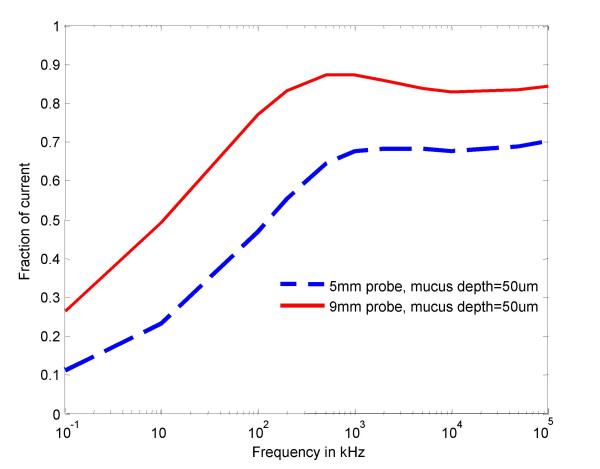
Background: We sought to compare uterine cervical electrical impedance spectroscopy measurements employing two probes of different sizes, and to employ a finite element model to predict and compare the fraction of electrical current derived from subepithelial stromal tissue.
Methods: Cervical impedance was measured in 12 subjects during early pregnancy using 2 different sizes of the probes on each subject.
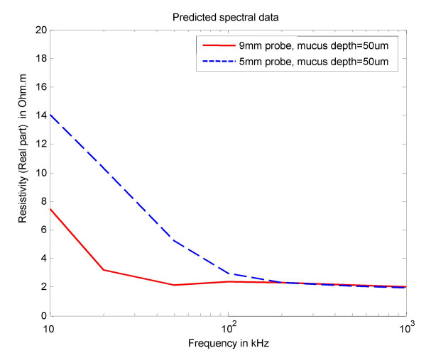
Results: Mean cervical resistivity was significantly higher (5.4 vs. 2.8 Omegam; p < 0.001) with the smaller probe in the frequency rage of 4-819 kHz. There was no difference in the short-term intra-observer variability between the two probes. The cervical impedance measurements derived in vivo followed the pattern predicted by the finite element model.
Conclusion: Inter-electrode distance on the probes for measuring cervical impedance influences the tissue resistivity values obtained. Determining the appropriate probe size is necessary when conducting clinical studies of resistivity of the cervix and other human tissues.
Figures

Similar articles
-
Reproducibility and repeatability of measuring the electrical impedance of the pregnant human cervix-the effect of probe size and applied pressure.Biomed Eng Online. 2009 Jun 17;8:10. doi: 10.1186/1475-925X-8-10. Biomed Eng Online. 2009. PMID: 19534806 Free PMC article.
-
Study of the optimum level of electrode placement for the evaluation of absolute lung resistivity with the Mk3.5 EIT system.Physiol Meas. 2006 May;27(5):S129-37. doi: 10.1088/0967-3334/27/5/S11. Epub 2006 Apr 20. Physiol Meas. 2006. PMID: 16636404 Clinical Trial.
-
Electrical impedance spectroscopy of the cervix in non-pregnant and pregnant women.Eur J Obstet Gynecol Reprod Biol. 2006 Dec;129(2):145-9. doi: 10.1016/j.ejogrb.2005.12.029. Epub 2006 Mar 6. Eur J Obstet Gynecol Reprod Biol. 2006. PMID: 16517044
-
Body fluid volumes measurements by impedance: A review of bioimpedance spectroscopy (BIS) and bioimpedance analysis (BIA) methods.Med Eng Phys. 2008 Dec;30(10):1257-69. doi: 10.1016/j.medengphy.2008.06.009. Epub 2008 Aug 3. Med Eng Phys. 2008. PMID: 18676172 Review.
-
Electrode-tissues interface: modeling and experimental validation.Biomed Mater. 2007 Mar;2(1):S7-S15. doi: 10.1088/1748-6041/2/1/S02. Epub 2007 Mar 2. Biomed Mater. 2007. PMID: 18458423 Review.
Cited by
-
In vivo Raman spectroscopy monitors cervical change during labor.Am J Obstet Gynecol. 2022 Aug;227(2):275.e1-275.e14. doi: 10.1016/j.ajog.2022.02.019. Epub 2022 Feb 19. Am J Obstet Gynecol. 2022. PMID: 35189092 Free PMC article.
-
A Narrative Review on the Clinical Utility of Electrical Impedance Spectroscopy for Diagnosing High-Grade Cervical Intraepithelial Neoplasia.Cureus. 2024 Jun 6;16(6):e61784. doi: 10.7759/cureus.61784. eCollection 2024 Jun. Cureus. 2024. PMID: 38975502 Free PMC article. Review.
-
Towards BirthAlert--A Clinical Device Intended for Early Preterm Birth Detection.IEEE Trans Biomed Eng. 2013 Dec;60(12):3484-93. doi: 10.1109/TBME.2013.2272601. Epub 2013 Jul 23. IEEE Trans Biomed Eng. 2013. PMID: 23893706 Free PMC article.
-
Tissue Characterization Using an Electrical Bioimpedance Spectroscopy-Based Multi-Electrode Probe to Screen for Cervical Intraepithelial Neoplasia.Diagnostics (Basel). 2021 Dec 14;11(12):2354. doi: 10.3390/diagnostics11122354. Diagnostics (Basel). 2021. PMID: 34943591 Free PMC article.
-
New techniques in evaluation of the cervix.Semin Perinatol. 2017 Dec;41(8):477-484. doi: 10.1053/j.semperi.2017.08.006. Semin Perinatol. 2017. PMID: 29191290 Free PMC article. Review.
References
-
- Brown BH, Milnes P, Abdul S, Tidy JA. Detection of cervical intraepithelial neoplasia using impedance spectroscopy: a prospective study. British Journal of Obstetrics and Gynaecology. 2005;112:802–806. - PubMed
Publication types
MeSH terms
LinkOut - more resources
Full Text Sources
Other Literature Sources
Medical
