. 2007 Mar;94(3):163-74.
doi: 10.1016/j.jip.2006.10.003. Epub 2006 Nov 27.

Infection of Ixodes scapularis ticks with Rickettsia monacensis expressing green fluorescent protein: a model system

Affiliations

Infection of Ixodes scapularis ticks with Rickettsia monacensis expressing green fluorescent protein: a model system

Gerald D Baldridge et al. J Invertebr Pathol. 2007 Mar.

Abstract

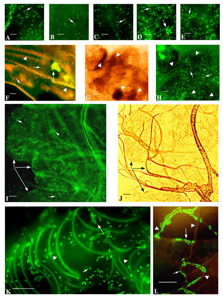
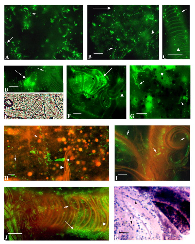
Ticks (Acari: Ixodidae) are ubiquitous hosts of rickettsiae (Rickettsiaceae: Rickettsia), obligate intracellular bacteria that occur as a continuum from nonpathogenic arthropod endosymbionts to virulent pathogens of both arthropod vectors and vertebrates. Visualization of rickettsiae in hosts has traditionally been limited to techniques utilizing fixed tissues. We report epifluorescence microscopy observations of unfixed tick tissues infected with a spotted fever group endosymbiont, Rickettsia monacensis, transformed to express green fluorescent protein (GFP). Fluorescent rickettsiae were readily visualized in tick tissues. In adult female, but not male, Ixodes scapularis infected by capillary feeding, R. monacensis disseminated from the gut and infected the salivary glands that are crucial to the role of ticks as vectors. The rickettsiae infected the respiratory tracheal system, a potential dissemination pathway and possible infection reservoir during tick molting. R. monacensis disseminated from the gut of capillary fed I. scapularis nymphs and was transstadially transmitted to adults. Larvae, infected by immersion, transstadially transmitted the rickettsiae to nymphs. Infected female I. scapularis did not transovarially transmit R. monacensis to progeny and the rickettsiae were not horizontally transmitted to a rabbit or hamsters. Survival of infected nymphal and adult I. scapularis did not differ from that of uninfected control ticks. R. monacensis did not disseminate from the gut of capillary fed adult female Amblyomma americanum (L.), or adult Dermacentor variabilis (Say) ticks of either sex. Infection of I. scapularis with R. monacensis expressing GFP provides a model system allowing visualization and study of live rickettsiae in unfixed tissues of an arthropod host.


Figures

Figure 1
Figure 1
Epifluorescence imaging of GFP transformant Rickettsia monacensis (Rmona658) in Amblyomma americanum, Dermacentor variabilis and Ixodes scapularis adult female ticks. Scale bar at lower left in each image indicates 10 μm and small arrows indicate Rmona658. (A) One week PCF Amblyomma gut tissue. (B) Three week PCF Amblyomma gut tissue. (C) One week PCF Dermacentor gut tissue. (D) Four week PCF Dermacentor gut tissue. Large arrow indicates dense cluster of Rmona658. (E) One week PCF Ixodes gut tissue. (F) Two week PCF Ixodes gut tissue imaged in red fluorescence channel with Rmona658 imaged in green channel. Arrowheads indicate trachea associated with gut. Large arrow indicates dense cluster of Rmona658 associated with trachea. (G and H) Visible light and epifluorescence images, respectively, of two week PCF Ixodes gut. Arrowheads indicate dark areas of dense hematin content. (I and J) Epifluorescence and visible light images, respectively, of two week PCF Ixodes salivary gland. Large arrows indicate distal edges of salivary gland acini. (K) Ixodes trachea with branch point at right side of image. Large arrow indicates cluster of doublet cell Rmona658. Arrowheads indicate taenidia. (L) Ixodes tracheoles (arrowheads) imaged in red fluorescence channel and associated with single and doublet cell (large arrow) forms of Rmona658 imaged in green fluorescence channel.
Figure 2
Figure 2
Imaging of GFP fluorescent Rmona658 in I. scapularis ticks infected as nymphs by capillary feeding. Scale bars at lower left in each image indicate 10 μm and small arrows indicate Rmona658. (A) One week PCF nymph gut tissue. Large arrow indicates chain of multiple Rmona658 cells. (B) Adult female salivary gland 4 days post-eclosion. Large arrow indicates distal edge of acini and arrowhead indicates salivary gland duct. (C) Adult female salivary gland duct 4 days post-eclosion. Arrowhead indicates junction of main and lateral salivary ducts. (D and E) Fluorescence and visible light images, respectively, of heavily infected adult female salivary gland duct 4 days post-eclosion. Large arrow indicates dense cluster of Rmona658. (F) Heavily infected complex of trachea and gut tissue from 4 day post-eclosion adult female. Large arrow indicates dense mass of Rmona658. Arrowhead indicates hematin granule. (G) Adult male gut tissue at 6 months post-eclosion. Arrowhead indicates hematin granule. (H) Adult female gut tissue imaged in red fluorescence channel at 6 months post-eclosion. Large arrow indicates dense cluster of Rmona658 imaged in green channel. Arrowhead indicates branching trachea associated with gut. (I) Adult female trunk and lateral tracheae imaged in red fluorescence channel at 6 months post-eclosion with Rmona658 imaged in green channel. (J) Heavily infected adult female trachea imaged in red fluorescence channel at 6 months post-eclosion. Large arrow indicates dense cluster of Rmona658 imaged in green channel. Arrowhead indicates taenidia. Infected tracheoles appear above scale bar. (K) Giemsa-stained adult ovarian tissue smear infected with rickettsia-like bacteria at 6 months post-eclosion.
Figure 3
Figure 3
PCR and Southern blot detection of Rmona658 transstadial transmission from I. scapularis nymphs to adults. (A) PCR amplification of a 460 bp tick mitochondrial 16S rRNA gene product from individual adult male (lanes 1 – 7) and female (lanes 8 – 14) tick tissue extracts. (B) Nested PCR amplification of an Rmona658-specific 400 bp GFP gene product from the same tick extracts as in A. (C) Southern blot detection of a 720 bp GFP gene 1st round PCR product from the same tick extracts as in A and B. Lanes L = 100 bp ladder with 500 bp band indicated at left. Lanes −C = no template and uninfected tick control extract in panel A and panels B and C, respectively. Lanes +C = plasmid template positive control.
Figure 4
Figure 4
PCR and Southern blot detection of Rmona658 transstadial transmission from I. scapularis larvae to nymphs. (A) PCR amplification of a 460 bp tick mitochondrial 16S rRNA gene product from individual nymphal tick tissue extracts (lanes 1 – 18). (B) Nested PCR amplification of an Rmona658-specific 355 bp CAT gene product from the same tissue extracts. (C) Southern blot detection of a 660 bp CAT gene 1st round PCR product from the same tick extracts as in A and B. Lanes L, −C and +C as in Fig. 3.
Figure 5
Figure 5
PCR detection of SFG rickettsiae in nymphal progeny of Rmona658-fed adult female I. scapularis. Upper and lower panels: amplification of a 532bp SFG rickettsial rompA gene product from DNA extracts of 6 pools of 5 nymphs each (lanes P1 to P6) and from 20 individual nymphs (lanes N1 to N20). Lanes L, −C and +C as in Fig. 3.

References

    1. Adams JR, Schmidtmann ET, Azad AF. Infection of colonized fleas Ctenocephalides felis (Bouche), with a rickettsia-like microorganism. Am J Trop Med Hyg. 1990;43:400–409. - PubMed
    1. Azad AF, Beard CB. Rickettsial pathogens and their arthropod vectors. Emerg Infect Dis. 1998;4:179–186. - PMC - PubMed
    1. Balashov YS. Bloodsucking ticks (Ixodoidea) - vectors of diseases of man and animals. Misc Pub Ent Soc Amer. 1972;8:5.
    1. Baldridge GD, Burkhardt NY, Simser JA, Kurtti TJ, Munderloh UG. Sequence and expression analysis of the rompA gene of Rickettsia peacockii, an endosymbiont of the Rocky Mountain Wood Tick, Dermacentor andersoni. Appl Environ Microbiol. 2004;70:6628–6636. - PMC - PubMed
    1. Baldridge GD, Burkhardt N, Herron MJ, Kurtti TJ, Munderloh UG. Analysis of fluorescent protein expression in transformants of Rickettsia monacensis, an obligate intracellular tick symbiont. Appl Environ Microbiol. 2005;71:2095–2105. - PMC - PubMed

Publication types

Substances

LinkOut - more resources