. 2007 Jan 12;1128(1):21-32.
doi: 10.1016/j.brainres.2006.10.056. Epub 2006 Nov 28.

Analysis of transcriptional modulation of the presenilin 1 gene promoter by ZNF237, a candidate binding partner of the Ets transcription factor ERM

Affiliations

Analysis of transcriptional modulation of the presenilin 1 gene promoter by ZNF237, a candidate binding partner of the Ets transcription factor ERM

Martine Pastorcic et al. Brain Res. .

Abstract

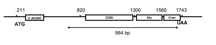
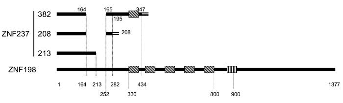
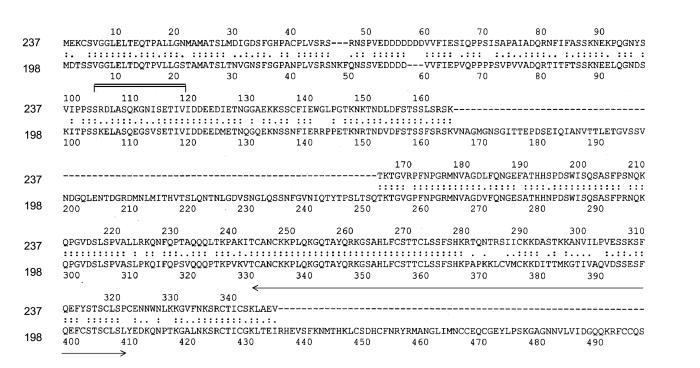
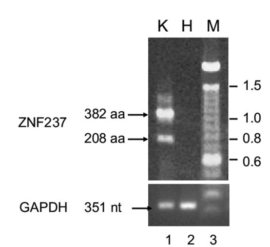
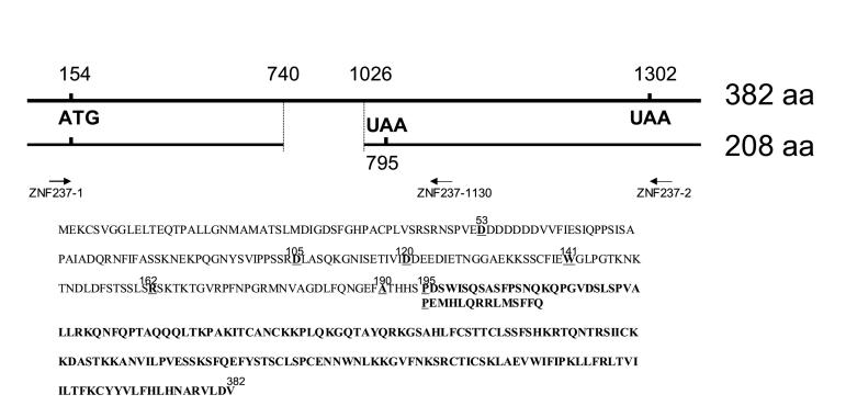
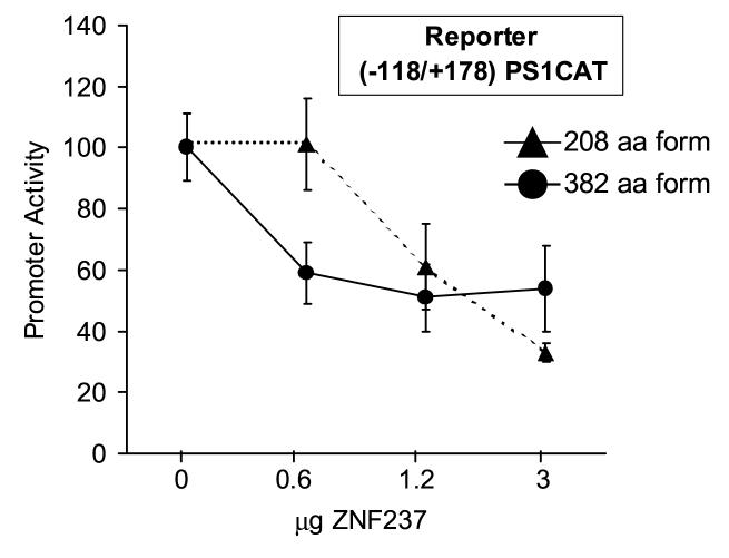
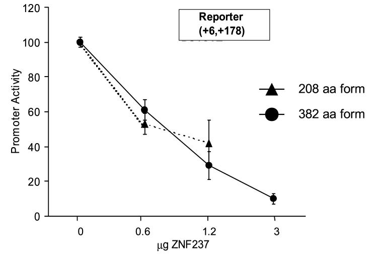
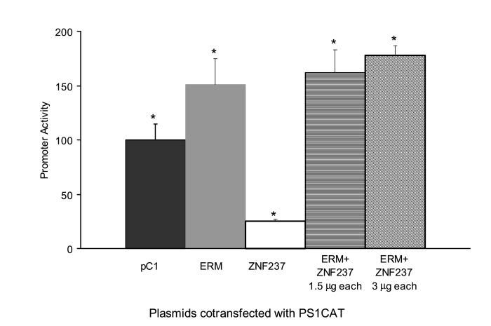
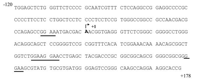
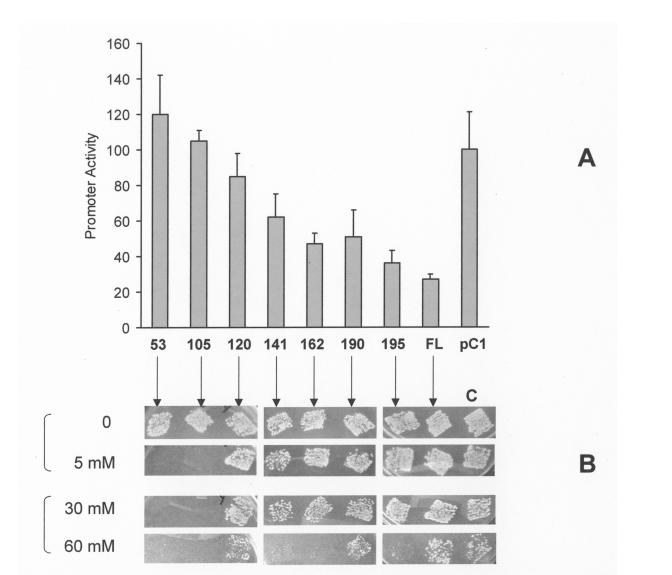
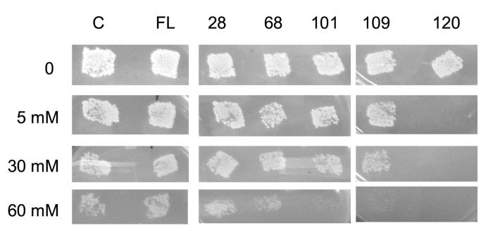
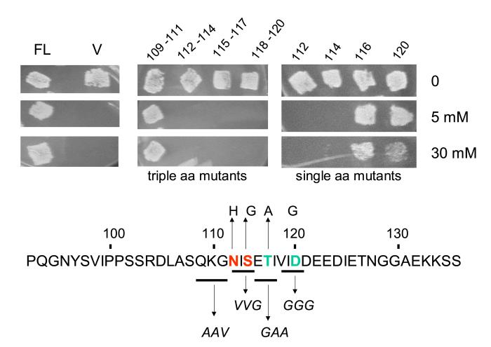
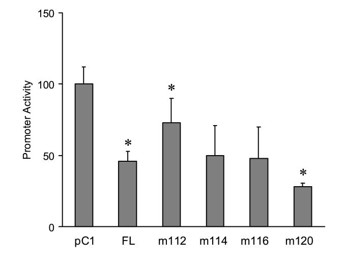
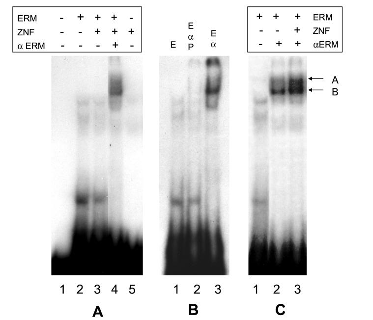
DNA sequences required for the expression of the human presenilin 1 (PS1) gene have been identified between -118 and +178 flanking the major initiation site (+1) mapped in SK-N-SH cells. Several Ets sites are located both upstream as well as downstream from the +1 site, including an Ets motif present at -10 that controls 90% of transcription in SK-N-SH cells. However, in SH-SY5Y cells, transcription initiates further downstream and requires an alternative set of promoter elements including a +90 Ets motif. Ets2, ER81, ERM and Elk1 were identified by yeast one-hybrid selection in a human brain cDNA library using the -10 Ets motif as a bait. We have shown that ERM recognizes specifically Ets motifs on the PS1 promoter located at -10 as well as downstream at +90, +129 and +165 and activates PS1 transcription with promoter fragments whether or not they contain the -10 Ets site. We have now searched for ERM interacting proteins by yeast two-hybrid selection in a human brain cDNA library using the C-terminal 415 amino acid of ERM as a bait. One of the interacting proteins was ZNF237, a member of the MYM gene family. It is widely expressed in different tissues in eukaryotes under several forms derived by alternative splicing, including a large 382 amino acid form containing a single MYM domain, and 2 shorter forms of 208 and 213 amino acids respectively that do not. We show that both the 382 as well as the 208 amino acid forms are expressed in SK-N-SH cells but not in SH-SY5Y cells. Both forms interact with ERM and repress the transcription of PS1 in SH-SY5Y cells. The effect of both C-terminal and N-terminal deletions indicates that the N-terminal 120 amino acid region is required for interaction with ERM in yeast, and furthermore single amino acid mutations show that residues 112 and 114 play an important role. The repression of transcription in SH-SY5Y cells also appears to require the N-terminal potion of ZNF237 and was affected by mutation of the amino acid 112. Data from electrophoretic mobility shift assays indicate that ERM and possibly ZNF237 interact with a fragment of the PS1 promoter.


Figures

Figure 1
Figure 1
ERM protein domains included in the yeast two-hybrid bait. The linear sequence of the cDNA for ERM is annotated according to GI: 33873571. The translation starts at nucleotide 211 and stops at position 1743. The domains conserved within the PEA3 family are boxed, including the N-terminal alpha-helical acidic domain contained within the first 72 amino acids, the Ets domain, the central inhibitory DNA-binding domain (CIDD) and the C-terminal domain. The fragment of cDNA included in the bait is indicated by arrows.
Figure 2
Figure 2
Various ZNF237 proteins and their homologies. A. Homologous regions between ZNF237 and ZNF198. The 382 amino acid major ZNF 237 (GI: 6010116) form is aligned with the 2 minor 208 (GI: 6010118) and 213 (GI: 6010120) amino acid forms derived by alternative splicing as well as with ZNF198 (GI: 22137330) in order to display homologous segments. Black lines show homologous areas. Double lines mark heterologous regions: amino acid 195 to 208 of ZNF237 (208 aa form) are absent in ZNF237 (382aa form) or ZNF198. The triple line at amino acid 347 in ZNF237 (382 aa form) marks a region of divergence with ZNF198. The MYM motifs are indicated by boxes striped in diagonal, the proline rich region of ZNF198 is shown by a box with vertical stripes. The amino acid positions on the respective proteins are indicated by numbers. B. Amino acid sequences alignment of ZNF237 and ZNF198 . The N-terminal amino acids 1 to 347 of ZNF237 were aligned with the homologous region in the N-terminal of ZNF198. The area including an MYM domain is underlined with arrows. The protein region where mutations eliminate binding to ERM by yeast-two hybrid assays is indicated by brackets. Dotted lines indicate residues in ZNF198 that are absent in ZNF 237. Dots represent amino acids which are conserved between ZNF237 and ZNF198.
Figure 2
Figure 2
Various ZNF237 proteins and their homologies. A. Homologous regions between ZNF237 and ZNF198. The 382 amino acid major ZNF 237 (GI: 6010116) form is aligned with the 2 minor 208 (GI: 6010118) and 213 (GI: 6010120) amino acid forms derived by alternative splicing as well as with ZNF198 (GI: 22137330) in order to display homologous segments. Black lines show homologous areas. Double lines mark heterologous regions: amino acid 195 to 208 of ZNF237 (208 aa form) are absent in ZNF237 (382aa form) or ZNF198. The triple line at amino acid 347 in ZNF237 (382 aa form) marks a region of divergence with ZNF198. The MYM motifs are indicated by boxes striped in diagonal, the proline rich region of ZNF198 is shown by a box with vertical stripes. The amino acid positions on the respective proteins are indicated by numbers. B. Amino acid sequences alignment of ZNF237 and ZNF198 . The N-terminal amino acids 1 to 347 of ZNF237 were aligned with the homologous region in the N-terminal of ZNF198. The area including an MYM domain is underlined with arrows. The protein region where mutations eliminate binding to ERM by yeast-two hybrid assays is indicated by brackets. Dotted lines indicate residues in ZNF198 that are absent in ZNF 237. Dots represent amino acids which are conserved between ZNF237 and ZNF198.
Figure 3
Figure 3
RNA and protein structure of ZNF237 major 382 amino acid form and 208 amino acid minor form. A. RT-PCR with total cellular RNA from SK-N-SH cells (lane 1) or SH-SY5Y (lane 2) with primers ZNF237-1 and ZNF237-1130. Molecular weight markers were run alongside (lane 3) and fragment size is indicated in kb on the right. Arrows show the fragments corresponding the major ZNF237 specie of 382 amino acids and the minor 208 amino acid form. Control PCR reactions to the human GAPDH gene were performed with each of the cDNAs. The expected 351 nt GAPDH product is indicated by an arrow. Primers used for the GAPDH gene were: sense
  1. 5′-AGCAATGCCTCCTGCACCACCAAC-3′

  2. and antisense 5′-CTGCTTCACCACCTTCTTGATG-3′.

B. Top: the homologous regions of the cDNAs from the 382 and 208 amino acid forms are aligned. The numbers indicate the nucleotide positions according to GI: 6010116. The start (ATG) and stop (UAA) of translation are indicated. The position of primers used to identify and clone the cDNAs by RT-PCR is indicated by arrows. Bottom: the amino acid sequence of both proteins is summarized: amino acid 1 to 195 are common to both 382 and 208 species. The divergent C-terminal regions downstream from 195 are indicated in bold. The numbers near the amino acid sequence indicate the position of the underlined residues.
Figure 3
Figure 3
RNA and protein structure of ZNF237 major 382 amino acid form and 208 amino acid minor form. A. RT-PCR with total cellular RNA from SK-N-SH cells (lane 1) or SH-SY5Y (lane 2) with primers ZNF237-1 and ZNF237-1130. Molecular weight markers were run alongside (lane 3) and fragment size is indicated in kb on the right. Arrows show the fragments corresponding the major ZNF237 specie of 382 amino acids and the minor 208 amino acid form. Control PCR reactions to the human GAPDH gene were performed with each of the cDNAs. The expected 351 nt GAPDH product is indicated by an arrow. Primers used for the GAPDH gene were: sense
  1. 5′-AGCAATGCCTCCTGCACCACCAAC-3′

  2. and antisense 5′-CTGCTTCACCACCTTCTTGATG-3′.

B. Top: the homologous regions of the cDNAs from the 382 and 208 amino acid forms are aligned. The numbers indicate the nucleotide positions according to GI: 6010116. The start (ATG) and stop (UAA) of translation are indicated. The position of primers used to identify and clone the cDNAs by RT-PCR is indicated by arrows. Bottom: the amino acid sequence of both proteins is summarized: amino acid 1 to 195 are common to both 382 and 208 species. The divergent C-terminal regions downstream from 195 are indicated in bold. The numbers near the amino acid sequence indicate the position of the underlined residues.
Figure 4
Figure 4
Inhibition of PS1 transcription by ZNF237. SH-SY5Y cells were transfected with 6 μg of (-118,+178) PS1CAT (A) or (+6,+178) PS1CAT (B) reporter together with increasing amounts of ZNF237 major 382 (circles) or 208 (triangles) amino acid forms. The total amount of (ZNF237+pC1) was kept constant at 3 μg. Promoter activity in different samples was standardized using the amount of protein present in the cellular extracts as an internal control. Each experiment was repeated three times, with at the minimum triplicate tests of each construct combination. (C) (-118,+176) PS1 CAT (5 μg) was cotransfected with expression vectors for ERM or ZNF237 (208 aa form) (3 μg), or a mixture of ERM and ZNF237 (1.5 μg each or 3 μg) with a minimum triplicate test of each construct combination. The symbol (*) indicates differences significant with p<0.05 bt t-test/ANOVA between the pC1 control as compared to each combination of plasmid (s) tested. (D) DNA sequence of the PS1 promoter region used in transfection experiments. The homologies to the Ets motif are underlined. The major transcription initiation site is indicated at +1.
Figure 4
Figure 4
Inhibition of PS1 transcription by ZNF237. SH-SY5Y cells were transfected with 6 μg of (-118,+178) PS1CAT (A) or (+6,+178) PS1CAT (B) reporter together with increasing amounts of ZNF237 major 382 (circles) or 208 (triangles) amino acid forms. The total amount of (ZNF237+pC1) was kept constant at 3 μg. Promoter activity in different samples was standardized using the amount of protein present in the cellular extracts as an internal control. Each experiment was repeated three times, with at the minimum triplicate tests of each construct combination. (C) (-118,+176) PS1 CAT (5 μg) was cotransfected with expression vectors for ERM or ZNF237 (208 aa form) (3 μg), or a mixture of ERM and ZNF237 (1.5 μg each or 3 μg) with a minimum triplicate test of each construct combination. The symbol (*) indicates differences significant with p<0.05 bt t-test/ANOVA between the pC1 control as compared to each combination of plasmid (s) tested. (D) DNA sequence of the PS1 promoter region used in transfection experiments. The homologies to the Ets motif are underlined. The major transcription initiation site is indicated at +1.
Figure 5
Figure 5
Effect of C-terminal deletions on the inhibition of transcription by ZNF237 and on the binding to ERM. A. Effect of C-terminal deletions on the inhibition of transcription by ZNF237. SH-SY5Y cells were transfected with 6 μg of the (-118, +178)PS1CAT reporter together with 5 μg of ZNF237 full length or each C-terminal deletion with the indicated end-point ( position 53, 105, 120, 141, 162, 190, 195, or the full length 208 amino acid form) or the empty pC1 vector. Promoter activity in different samples was standardized using the amount of protein present in the cellular extracts as an internal control. Each experiment was repeated three times, with a minimum of n=4 for each data point. B. Identification of ZNF237 domains interacting with ERM by yeast two-hybrid assay. The same C-terminal deletions of ZNF237 were introduced into pACT2 and transformed into AH109 pretransformed with the Gal4BD-ERM fusion bait. The ability of the mutants to promote growth on SD medium lacking Tryptophane, Leucine or Histidine and including increasing amounts of 3-AT (indicated alongside on the left) was compared after 4 days (0 and 5 mM 3-AT) and after 5 days (30 and 60 mM 3-AT). Patches from the series C were transformants with the original cDNA from ZNF237 obtained when screening the library that included most of the 382 amino acid form except for the first 24 amino acids.
Figure 6
Figure 6
Effect of N-terminal deletions on the interaction of ZNF237 with ERM: yeast two-hybrid assay. The N-terminal deletions of ZNF237 to amino acids 28, 68, 101, 109 and 120 were introduced into pACT2 and transformed into AH109 pretransformed with the Gal4BD-ERM fusion bait. The ability of the mutants to promote growth on SD medium lacking Tryptophane, Leucine or Histidine and including increasing amounts of 3-AT (indicated alongside on the left) was compared after 4 days (0 and 5 mM 3-AT) and after 5 days (30 and 60 mM 3-AT). Patches from the series C were transformants with the original cDNA from ZNF237 obtained when screening the library that included most of the 382 amino acid form except for the first 24 amino acids. FL indicates the intact 208 amino acid form fused to the pACT2 polylinker and Gal4AD at amino acid 4.
Figure 7
Figure 7
Effect of selective amino acid mutations in ZNF237 on its binding to ERM and the inhibition of transcription of PS1. A. Effect of amino acid mutations of ZNF237 on its interaction with ERM in yeast two-hybrid assays. Either triple mutants or single amino acid mutants of the 208 amino acid form of ZNF237 were introduced into pACT2 and transformed into AH109 pretransformed with the Gal4BD-ERM fusion bait. The ability of the mutants to promote growth on SD medium lacking Tryptophane, Leucine or Histidine and including 0, 5 and 30 mM 3-AT (indicated alongside on the right) was compared after 5 days to the wild type (FL) and the empty pACT2 (V). The amino acid sequence of the corresponding area of ZNF237 is indicated together with the amino acids mutated and the residues that were substituted. B. Effect of single amino acid mutations on the inhibition of PS1 transcription by ZNF237. SH-SY5Y cells were transfected with 5 μg of the (-118, +178)PS1CAT reporter together with 6 μg of ZNF237 wild type 208 amino acid form or each of the mutants at single amino acid position 112, 114, 116 and 120 respectively. Promoter activity was standardized using the amount of protein present in the cellular extracts. Activity in the presence of the empty pC1 control vector was considered 100%. Each experiment was repeated three times, with a minimum of n=4 for each data point. The symbol (*) indicates a significant difference with p<0.05 by t-test/ANOVA between the full length protein and m112 as well as between the full length protein and m120.
Figure 7
Figure 7
Effect of selective amino acid mutations in ZNF237 on its binding to ERM and the inhibition of transcription of PS1. A. Effect of amino acid mutations of ZNF237 on its interaction with ERM in yeast two-hybrid assays. Either triple mutants or single amino acid mutants of the 208 amino acid form of ZNF237 were introduced into pACT2 and transformed into AH109 pretransformed with the Gal4BD-ERM fusion bait. The ability of the mutants to promote growth on SD medium lacking Tryptophane, Leucine or Histidine and including 0, 5 and 30 mM 3-AT (indicated alongside on the right) was compared after 5 days to the wild type (FL) and the empty pACT2 (V). The amino acid sequence of the corresponding area of ZNF237 is indicated together with the amino acids mutated and the residues that were substituted. B. Effect of single amino acid mutations on the inhibition of PS1 transcription by ZNF237. SH-SY5Y cells were transfected with 5 μg of the (-118, +178)PS1CAT reporter together with 6 μg of ZNF237 wild type 208 amino acid form or each of the mutants at single amino acid position 112, 114, 116 and 120 respectively. Promoter activity was standardized using the amount of protein present in the cellular extracts. Activity in the presence of the empty pC1 control vector was considered 100%. Each experiment was repeated three times, with a minimum of n=4 for each data point. The symbol (*) indicates a significant difference with p<0.05 by t-test/ANOVA between the full length protein and m112 as well as between the full length protein and m120.
Figure 8
Figure 8
ZNF237 interaction with the PS1 promoter. A. In vitro translated ZNF237 208 amino acid form and full length ERM were incubated together with a labeled probe containing PS1 promoter sequences from -22 to +6 and in the presence of the goat polyclonal anti-ERM antibody sc-1955X (αERM). Added components are marked (+). B. ERM (E) was incubated with the PS1 probe (lane 1), with added anti-ERM antibody (lane 3), or with both the antibody and the control peptide used to raise the antibody (P) (lane 2). C. ERM was incubated with the PS1 promoter (lane 1), together with anti-ERM antibody and ZNF237 (lane 3), or a control aliquot of an in vitro translation reaction with the empty pCMV-Tag 2 vector as a template instead of ZNF237 (lane 2). Arrows indicate the position of specific DNA-protein complexes.

Similar articles

Cited by

References

    1. Baumann H, Kunapuli P, Tracy E, Cowell JK. The oncogenic fusion protein-tyrosine kinase ZNF198/fibroblast growth factor receptor-1 has signaling function comparable with interleukin-6 cytokine receptors. J Biol Chem. 2003;278:16198–16208. - PubMed
    1. Chen J, Deangelo DJ, Kutok JL, Williams IR, Lee BH, Wadleigh M, Duclos N, Cohen S, Adelsperger J, Okabe R, Coburn A, Galinsky I, Huntly B, Cohen PS, Meyer T, Fabbro D, Roesel J, Banerji L, Griffin JD, Xiao S, Fletcher JA, Stone RM, Gilliland DG. PKC412 inhibits the zinc finger 198-fibroblast growth factor receptor 1 fusion tyrosine kinase and is active in treatment of stem cell myeloproliferative disorder. Proc Natl Acad Sci U S A. 2004;101:14479–14484. - PMC - PubMed
    1. Chotteau-Lelievre A, Dolle P, Peronne V, Coutte L, de Launoit Y, Desbiens X. Expression patterns of the Ets transcription factors from the PEA3 group during early stages of mouse development. Mech Dev. 2001;108:191–195. - PubMed
    1. Chotteau-Lelievre A, Montesano R, Soriano J, Soulie P, Desbiens X, de Launoit Y. PEA3 transcription factors are expressed in tissues undergoing branching morphogenesis and promote formation of duct-like structures by mammary epithelial cells in vitro. Dev Biol. 2003;259:241–257. - PubMed
    1. Chyung JH, Raper DM, Selkoe DJ. Gamma-secretase exists on the plasma membrane as an intact complex that accepts substrates and effects intramembrane cleavage. J Biol Chem. 2005;280:4383–4392. - PubMed

Publication types

MeSH terms