. 2007 Feb;25(2):297-308.
doi: 10.1016/j.nbd.2006.09.016. Epub 2006 Nov 27.

Cardiac dysfunction in the R6/2 mouse model of Huntington's disease

Affiliations

Cardiac dysfunction in the R6/2 mouse model of Huntington's disease

Michael J Mihm et al. Neurobiol Dis. 2007 Feb.

Abstract

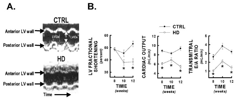
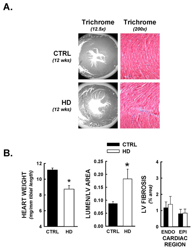
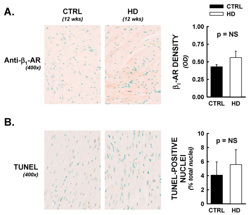
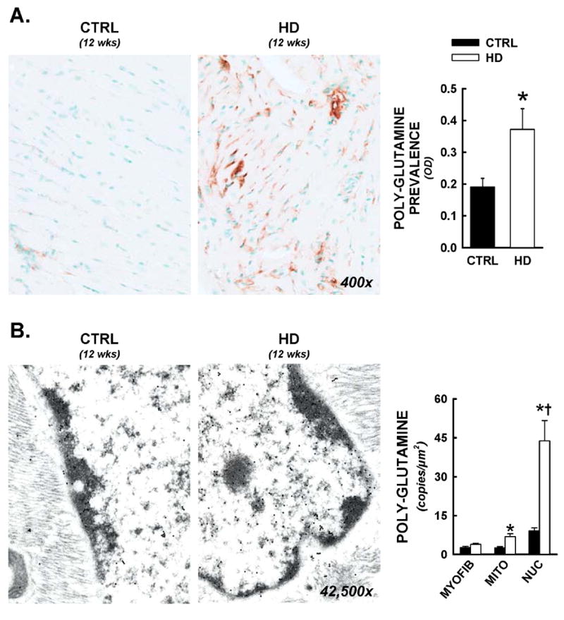
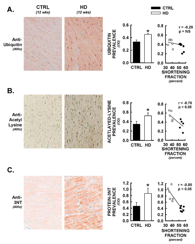
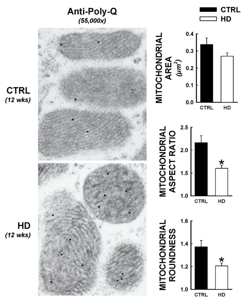
Recent evidence suggests that mutant huntingtin protein-induced energetic perturbations contribute to neuronal dysfunction in Huntington's disease (HD). Given the ubiquitous expression of huntingtin, other cell types with high energetic burden may be at risk for HD-related dysfunction. Early-onset cardiovascular disease is the second leading cause of death in HD patients; a direct role for mutant huntingtin in this phenomenon remains unevaluated. Here we tested the hypothesis that expression of mutant huntingtin is sufficient to induce cardiac dysfunction, using a well-described transgenic model of HD (line R6/2). R6/2 mice developed cardiac dysfunction by 8 weeks of age, progressing to severe failure at 12 weeks, assessed by echocardiography. Limited evidence of cardiac remodeling (e.g. hypertrophy, fibrosis, apoptosis, beta(1) adrenergic receptor downregulation) was observed. Immunogold electron microscopy demonstrated significant elevations in nuclear and mitochondrial polyglutamine presence in the R6/2 myocyte. Significant alterations in mitochondrial ultrastructure were seen, consistent with metabolic stress. Increased cardiac lysine acetylation and protein nitration were observed and were each significantly associated with impairments in cardiac performance. These data demonstrate that mutant huntingtin expression has potent cardiotoxic effects; cardiac failure may be a significant complication of this important experimental model of HD. Investigation of the potential cardiotropic effects of mutant huntingtin in humans may be warranted.


Figures

Figure 1
Figure 1
Echocardiographic evidence of decreased cardiac performance in HD mice. HD mice and littermate controls (CTRL) were serially assessed for systolic and diastolic cardiac performance at 8, 10, and 12 weeks of age by non-invasive echocardiography. Panel A) Representative M-mode images of control and HD cardiac left ventricles at 12 weeks of age. HD mice demonstrated significantly diminished anterior and posterior wall motion compared to littermate controls. Panel B) Selected systolic (LV fractional shortening, cardiac output) and diastolic (transmitral E/A ratio) cardiac performance indicators from HD mice and littermate controls at 8, 10, and 12 weeks. * = p<0.05 from control values at equivalent time point.
Figure 2
Figure 2
Limited evidence of cardiac structural remodeling in HD mice. Following functional analyses, hearts were prepared for histopathological studies, and stained with Masson’s Trichrome (Myocardium = red, nuclei = black, collagen/fibrosis = blue). All data are from animals sacrificed at 12 weeks of age. Panel A) Representative photomicrographs of Masson’s Trichrome staining at 12.5 and 200x magnifications. Myocardial cross-sectional areas from HD mice were smaller than littermate control (CTRL) hearts, and demonstrated equivalent and limited evidence of interstitial fibrosis. Panel B) Heart weights (normalized to tibial length) for HD mice were significantly decreased relative to littermate controls. HD hearts demonstrated significant LV chamber dilation, determined by lumenal/myocardial area ratios. No increases in interstitial fibrosis was detected, as both endocardial (inner 50% of myocardium) and epicardial (outer 50% of myocardium) Trichrome staining were under 2% of total tissue area. *, p<0.05 versus control values at equivalent time point.
Figure 3
Figure 3
Cardiac β1-adrenergic receptor densities and myocyte apoptotic events were not altered in HD mice. Following functional analyses, hearts were assessed for β1-adrenergic receptor densities and DNA nicks by immunohistochemical methods. All data are from animals sacrificed at 12 weeks of age. Panel A) Representative photomicrographs from anti- β1 adrenergic receptor labelling (400x). No significant differences were observed between HD hearts and littermate controls, by optical density analysis. Panel B) TUNEL staining was employed as an indirect marker of apoptotic events. TUNEL positive nuclei (brown nuclei) were counted by an automated digital imaging approach, and expressed as a percentage of DNAse positive control (maximal nuclear staining) from serial sections. Percentages of total nuclei that were TUNEL-positive were not significantly different between treatment groups.
Figure 4
Figure 4
Nuclear and mitochondrial residence of mutant huntingtin in R6/2 mice. Following functional analyses, cardiac tissues were prepared for both light microscopy and electron microscopy immunohistochemistry. All data are from animals sacrificed at 12 weeks of age. Panel A) Representative photomicrographs from anti-polyglutamine staining (antibody raised against poly-glutamine repeats >37, 400x). Increased prevalence of the expanded polyglutamine sequence was visualized in both cytosolic and nuclear spaces. Total cardiac polyglutamine prevalence was significantly elevated in HD hearts by optical density analysis. Panel B) Immunogold electron microscopy for intracellular distributions of mutant huntingtin. Immunohistochemistry was conducted as above, with a gold-colloid secondary antibody (10nm particles, visualized as electron dense circles under tunneling electron microscopic examination). Images of myocytes with longitudinal orientation were captured and nuclear, mitochondrial, and myofibrillar areas were delineated and calibrated, then positive copies for the expanded polyglutamine sequence were counted, and expressed per nuclear, mitochondrial, or myofibrillar area. Mean data is represented in histogram format. *, p<0.05 versus littermate control.
Figure 5
Figure 5
Alterations in protein post-translational modifications in HD hearts. Following functional analyses, hearts were assessed for protein-ubiquitin, lysine acetylation, and protein-nitrotyrosine formation by immunohistochemical methods. All data are from animals sacrificed at 12 weeks of age. Panel A) Representative photomicrographs from anti-ubiquitin staining (400x). Increases in cytosolic and nuclear staining were observed in HD hearts. These changes were not significantly correlated to alterations in fractional shortening in these same mice (Spearman’s non-parametric correlation). Panel B) Protein lysine-acetylation was assessed using a pan-antibody to acetylated-lysine. Nuclear as well as cytosolic increases in acetylated-lysine were observed in HD hearts, and optical densities for cardiac lysine-acetylation were significantly negatively correlated to LV fractional shortening in these same mice. Panel C) Protein-3-nitrotyrosine (protein-3NT) was also increased in cardiac cross-sections from HD mice relative to controls. Staining was predominantly observed in cardiac myocytes themselves and was widespread throughout the myocardium. Extent of cardiac protein-3NT formation was significantly correlated to alterations in fractional shortening in these same mice. *, p<0.05 versus littermate control.
Figure 6
Figure 6
Altered mitochondrial ultrastructure in HD hearts. Electron micrographs from HD and control hearts were assessed using quantitative measures for mitochondrial morphology, at 12 weeks of age. These assessments were made in the same electron micrographs as immunogold studies for the expanded polyglutamine sequence, such that electron dense circles indicate mutant huntingtin presence. Over 300 individual mitochondrion were assessed for areas, aspect ratio (defined as the length of the major mitochondrial axis divided by the minor mitochondrial axis, which converges to 1.0 for a perfect circle), and roundness (defined as: [(mitochondrial perimeter)2]/[2π*(mitochondrial area)], which converges to 1.0 for a perfect circle). *, p<0.05 versus littermate control.

References

    1. Andreassen OA, Ferrante RJ, Dedeoglu A, Beal MF. Lipoic acid improves survival in transgenic mouse models of Huntington’s disease. Neuroreport. 2001a;12:3371–3373. - PubMed
    1. Andreassen OA, Dedeoglu A, Ferrante RJ, Jenkins BG, Ferrante KL, Thomas M, Friedlich A, Browne SE, Schilling G, Borchelt DR, Hersch SM, Ross CA, Beal MF. Creatine increase survival and delays motor symptoms in a transgenic animal model of Huntington’s disease. Neurobiol Dis. 2001b;8:479–491. - PubMed
    1. Arenas J, Campos Y, Ribacoba R, Martin MA, Rubio JC, Ablanedo P, Cabello A. Complex I defect in muscle from patients with Huntington’s disease. Annals of Neurology. 1998;43:397–400. - PubMed
    1. Bates G, Harper P, Jones L, editors. Huntington’s Disease. 3. Oxford ; New York: Oxford University Press; 2002.
    1. Bates GP, Mangiarini L, Davies SW. Transgenic mice in the study of polyglutamine repeat expansion diseases. Brain Pathol. 1998;8:699–714. - PMC - PubMed

Publication types

MeSH terms