

Involvement of chloride channels in IGF-I-induced proliferation of porcine arterial smooth muscle cells
- PMID: 17126821
- PMCID: PMC1852543
- DOI: 10.1016/j.cardiores.2006.10.012
Involvement of chloride channels in IGF-I-induced proliferation of porcine arterial smooth muscle cells
Abstract
Objective: The existence of Cl- channels in vascular smooth muscle cells (VSMCs) has been increasingly investigated, but the biological functions are not yet clear. Insulin-like growth factor (IGF)-I affects proliferation and migration of VSMCs, and dysregulation of this axis may be involved in atherogenesis and intimal hyperplasia. We examined the effects of Cl- channel blockers on IGF-I-induced proliferation in porcine VSMCs. The siRNA approach was used to support the role of ClC-2, a member of the volume-regulated Cl- channel family, in cell proliferation of VSMCs.
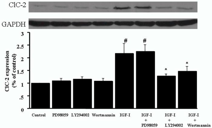
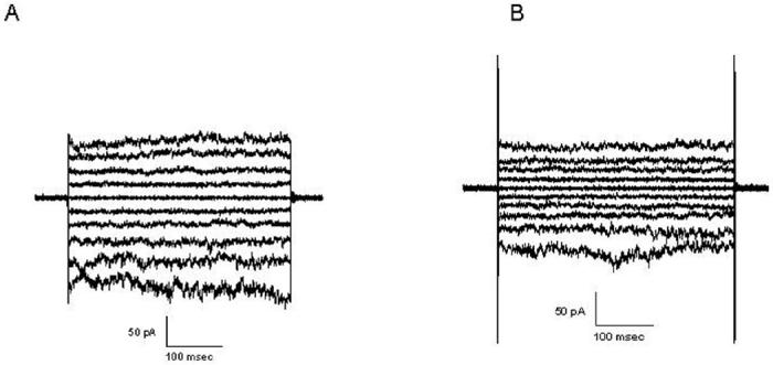
Methods and results: The IGF-I-induced VSMC proliferation was significantly suppressed by the Cl- channel blockers NPPB and IAA94 but not by DIDS. IGF-I-induced cell proliferation parallels a significant increase in the endogenous expression of ClC-2 mRNA and protein. Inhibitors of PI3-kinase, LY294002 and wortmannin, significantly attenuated the IGF-I-upregulated ClC-2 expression and cell proliferation. We observed ClC-2-like Cl- current, and this current was augmented by IGF-I. SiRNA specifically targeted to ClC-2 abolished IGF-I-induced cell proliferation.
Conclusion: Our data demonstrate that ClC-2 plays a role in IGF-1-induced regulation of VSMC proliferation in cardiovascular diseases.
Figures


Similar articles
-
Roles of phosphatidylinositol 3-kinase and mitogen-activated protein kinase pathways in stimulation of vascular smooth muscle cell migration and deoxyriboncleic acid synthesis by insulin-like growth factor-I.Endocrinology. 1999 Sep;140(9):4228-35. doi: 10.1210/endo.140.9.6980. Endocrinology. 1999. PMID: 10465296
-
Early Growth Response Protein-1 Expression by Insulin-Like Growth Factor-1 Requires ROS-Dependent Activation of ERK1/2 and PKB Pathways in Vascular Smooth Muscle Cells.J Cell Biochem. 2016 Jan;117(1):152-62. doi: 10.1002/jcb.25260. J Cell Biochem. 2016. PMID: 26084532
-
IGF-1 up-regulates K+ channels via PI3-kinase, PDK1 and SGK1.Pflugers Arch. 2002 Feb;443(4):625-34. doi: 10.1007/s00424-001-0741-5. Epub 2001 Nov 14. Pflugers Arch. 2002. PMID: 11907830
-
Transport and function of chloride in vascular smooth muscles.J Vasc Res. 2013;50(1):69-87. doi: 10.1159/000345242. Epub 2012 Nov 17. J Vasc Res. 2013. PMID: 23172353 Review.
-
The ClC-3 chloride channels in cardiovascular disease.Acta Pharmacol Sin. 2011 Jun;32(6):675-84. doi: 10.1038/aps.2011.30. Epub 2011 May 23. Acta Pharmacol Sin. 2011. PMID: 21602838 Free PMC article. Review.
Cited by
-
An angiotensin II- and NF-kappaB-dependent mechanism increases connexin 43 in murine arteries targeted by renin-dependent hypertension.Cardiovasc Res. 2010 Jul 1;87(1):166-76. doi: 10.1093/cvr/cvq031. Epub 2010 Jan 28. Cardiovasc Res. 2010. PMID: 20110337 Free PMC article.
-
Cl⁻ channels in smooth muscle cells.Pflugers Arch. 2014 May;466(5):861-72. doi: 10.1007/s00424-013-1357-2. Pflugers Arch. 2014. PMID: 24077695 Free PMC article. Review.
-
Chloride channelopathies of ClC-2.Int J Mol Sci. 2013 Dec 27;15(1):218-49. doi: 10.3390/ijms15010218. Int J Mol Sci. 2013. PMID: 24378849 Free PMC article. Review.
-
Inhibition of angiotensin II-induced cerebrovascular smooth muscle cell proliferation by LRRC8A downregulation through suppressing PI3K/AKT activation.Hum Cell. 2019 Jul;32(3):316-325. doi: 10.1007/s13577-019-00260-6. Epub 2019 May 24. Hum Cell. 2019. PMID: 31127489
-
ClC-2 knockdown prevents cerebrovascular remodeling via inhibition of the Wnt/β-catenin signaling pathway.Cell Mol Biol Lett. 2018 Jun 27;23:29. doi: 10.1186/s11658-018-0095-z. eCollection 2018. Cell Mol Biol Lett. 2018. PMID: 29988306 Free PMC article.
References
-
- Ross R. Atherosclerosis: an inflammatory disease. N Engl J Med. 1999;340:115–26. - PubMed
-
- Libby P. Coronary artery injury and the biology of atherosclerosis: inflammation, thrombosis, and stabilization. Am J Cardiol. 2000;86:3–8. - PubMed
-
- Delafontaine P. Insulin-like growth factor I and its binding proteins in the cardiovascular system. Cardiovasc Res. 1995;30:825–34. - PubMed
-
- Bayes-Genis A, Conover CA, Schwartz RS. The insulin-like growth factor axis: a review of atherosclerosis and restenosis. Circ Res. 2000;86:125–30. - PubMed
-
- Voets T, Szucs G, Droogmans G, Nilius B. Blockers of volume-activated Cl- currents inhibit endothelial cell proliferation. Pflugers Arch. 1995;431:132–134. - PubMed
