. 2007 Apr 10;360(2):305-21.
doi: 10.1016/j.virol.2006.10.028. Epub 2006 Nov 28.

Recruitment of activated IRF-3 and CBP/p300 to herpes simplex virus ICP0 nuclear foci: Potential role in blocking IFN-beta induction

Affiliations

Recruitment of activated IRF-3 and CBP/p300 to herpes simplex virus ICP0 nuclear foci: Potential role in blocking IFN-beta induction

Gregory T Melroe et al. Virology. .

Abstract

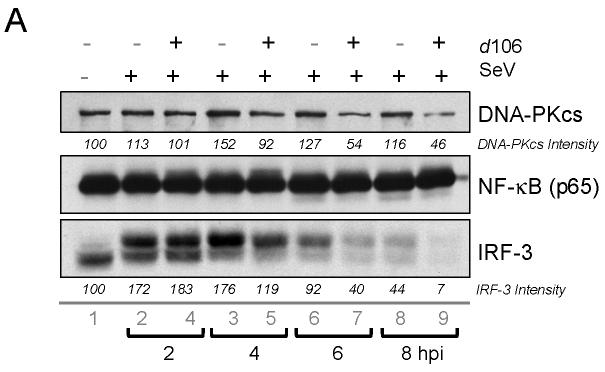
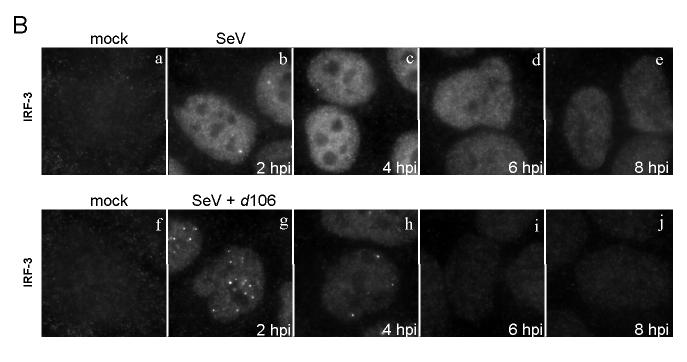
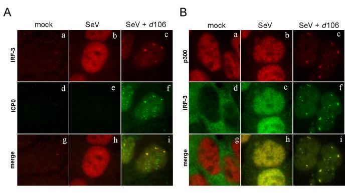
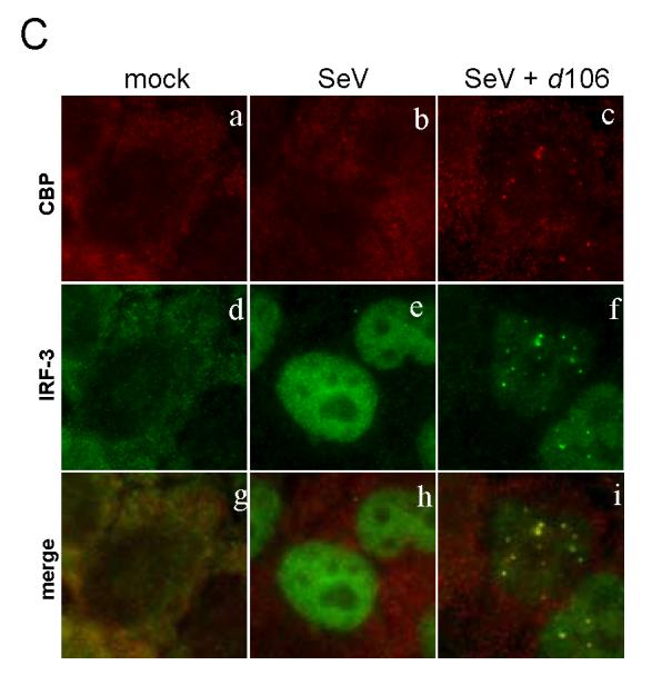
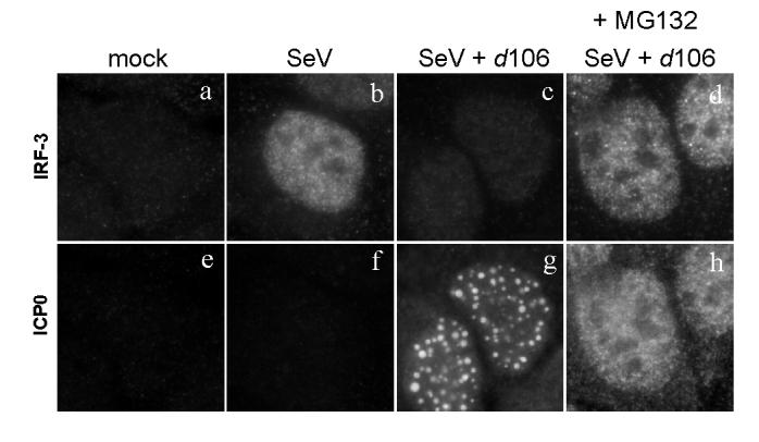
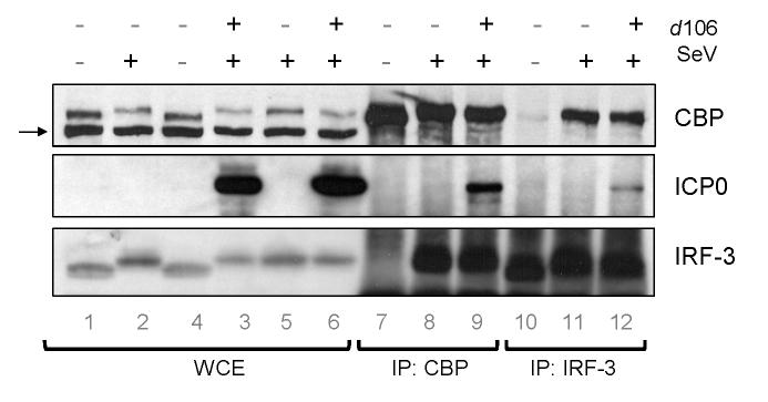
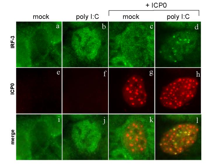
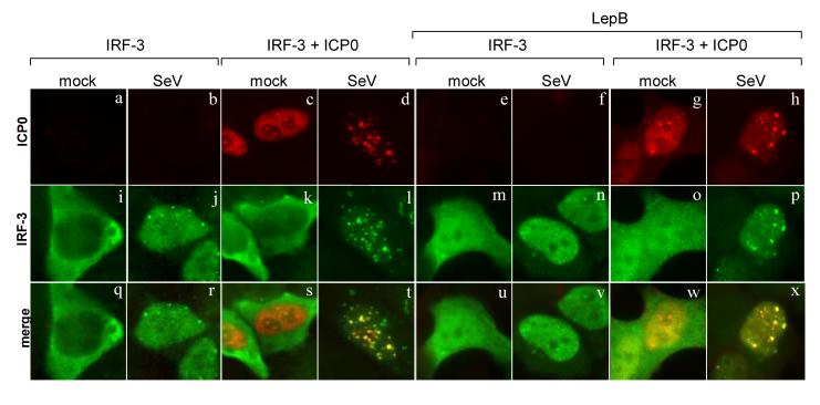
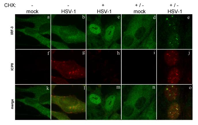
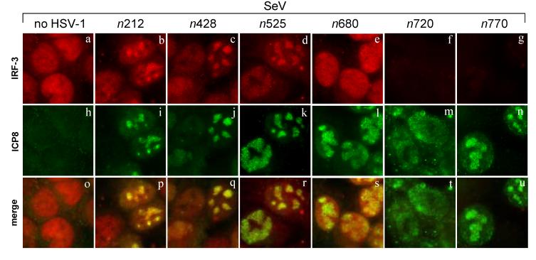
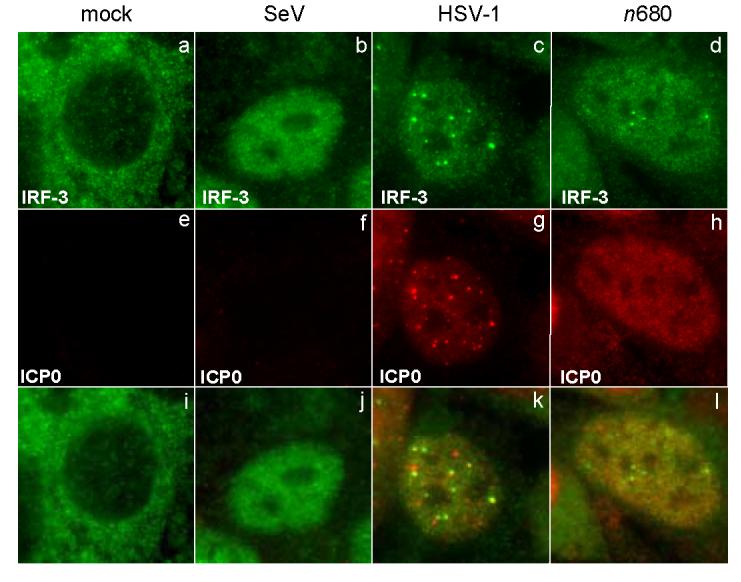
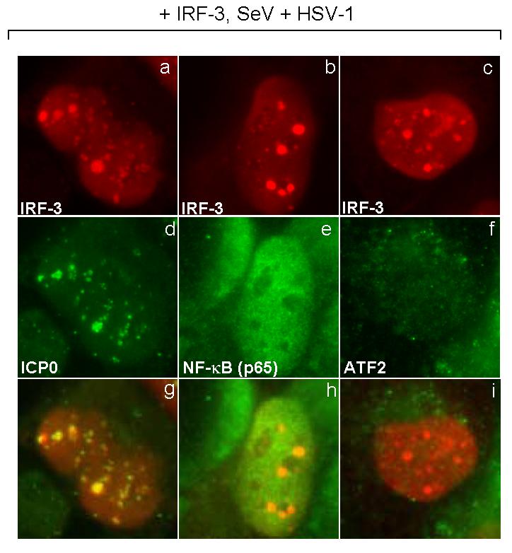
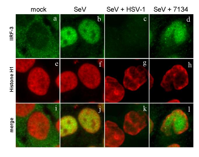
The host innate response to viral infection includes the production of interferons, which is dependent on the coordinated activity of multiple transcription factors. Herpes simplex virus 1 (HSV-1) has been shown to block efficient interferon expression by multiple mechanisms. We and others have demonstrated that HSV-1 can inhibit the transcription of genes promoted by interferon regulatory factor-3 (IRF-3), including interferon beta (IFN-beta), and that the immediate-early ICP0 protein is sufficient for this function. However, the exact mechanism by which ICP0 blocks IRF-3 activity has yet to be determined. Unlike some other viral proteins that inhibit IRF-3 activity, ICP0 does not appear to affect phosphorylation and dimerization of IRF-3. Here, we show that a portion of activated IRF-3 co-localizes with nuclear foci containing ICP0 at early times after virus infection. Co-localization to ICP0-containing foci is also seen with the IRF-3-binding partners and transcriptional co-activators, CBP and p300. In addition, using immunoprecipitation of infected cell lysates, we can immunoprecipitate a complex containing ICP0, IRF-3, and CBP. Thus we hypothesize that ICP0 recruits activated IRF-3 and CBP/p300 to nuclear structures, away from the host chromatin. This leads to the inactivation and accelerated degradation of IRF-3, resulting in reduced transcription of IFN-beta and an inhibition of the host response. Therefore, ICP0 provides an example of how viruses can block IFN-beta induction by sequestration of important transcription factors essential for the host response.


Figures

FIG. 1
FIG. 1
IRF-3 levels are reduced in d106-infected cells. (A) Western blot showing total levels of IRF-3 at different time points post-infection following exposure to SeV in the presence of absence of d106. Proteins in cell lysates were separated by SDS-PAGE and detected with antibodies towards DNA-PKcs, NF-kB (p65 subunit), and IRF-3. Individual bands corresponding to the proteins were scanned, and the intensity was quantified by computer analysis. The protein amounts were normalized to the amount present in mock-infected cells. The data presented are representative of multiple experiments. (B) Indirect immunofluorescence was used to determine the localization and relative amount of IRF-3 at different time points post-infection. After fixation, the cells were incubated with the SL12.1 antibody to IRF-3, followed by an Alexa Fluor 488 goat anti-mouse secondary antibody. Image exposure time was set by using the SeV-infected cells at 4 hpi and was kept constant in the other samples.
FIG. 1
FIG. 1
IRF-3 levels are reduced in d106-infected cells. (A) Western blot showing total levels of IRF-3 at different time points post-infection following exposure to SeV in the presence of absence of d106. Proteins in cell lysates were separated by SDS-PAGE and detected with antibodies towards DNA-PKcs, NF-kB (p65 subunit), and IRF-3. Individual bands corresponding to the proteins were scanned, and the intensity was quantified by computer analysis. The protein amounts were normalized to the amount present in mock-infected cells. The data presented are representative of multiple experiments. (B) Indirect immunofluorescence was used to determine the localization and relative amount of IRF-3 at different time points post-infection. After fixation, the cells were incubated with the SL12.1 antibody to IRF-3, followed by an Alexa Fluor 488 goat anti-mouse secondary antibody. Image exposure time was set by using the SeV-infected cells at 4 hpi and was kept constant in the other samples.
FIG. 2
FIG. 2
Change of IRF-3 localization in the presence of ICP0. HEC-1-B cells were infected with SeV in the presence or absence of d106. (A) The cells were fixed at 2.5 h post infection and stained with mouse anti-ICP0 and rabbit anti-IRF-3 antibodies with appropriate secondary antibodies. (B) The cells were stained with mouse anti-p300 and rabbit anti-IRF-3 antibodies with appropriate secondary antibodies. (C) The cells were stained with mouse anti-CBP and rabbit anti-IRF-3 antibodies with appropriate secondary antibodies.
FIG. 2
FIG. 2
Change of IRF-3 localization in the presence of ICP0. HEC-1-B cells were infected with SeV in the presence or absence of d106. (A) The cells were fixed at 2.5 h post infection and stained with mouse anti-ICP0 and rabbit anti-IRF-3 antibodies with appropriate secondary antibodies. (B) The cells were stained with mouse anti-p300 and rabbit anti-IRF-3 antibodies with appropriate secondary antibodies. (C) The cells were stained with mouse anti-CBP and rabbit anti-IRF-3 antibodies with appropriate secondary antibodies.
FIG. 3
FIG. 3
Effect of MG132 on nuclear accumulation of IRF-3. Infection of HEC-1-B cells was carried out as before, only this time the proteasomal inhibitor MG132 was added to select samples. MG132 (10μM) was added at 1 hour post infection and maintained in the medium until fixation at 6 hpi. The cells were stained with mouse anti-IRF-3 and rabbit anti-ICP0 antibodies with appropriate secondary antibodies.
FIG. 4
FIG. 4
Association of ICP0 with CBP and IRF-3 in infected cells. HEC-1-B cells were infected with the indicated viruses (SeV and/or d106) and harvested at 4 hpi. Shown is the Western blot analysis of proteins immunoprecipitated using polyclonal antibodies against CBP or IRF-3. Lanes 1-6 show a sample of the whole cell lysate (5% total). The arrow indicates a cellular protein band that cross-reacts with the CBP antibody. Lanes 7-9 are lanes with proteins immunoprecipitated using an anti-CBP antibody. Lanes 10-12 are lanes with proteins immunoprecipitated using an anti-IRF-3 antibody. The blot was probed with antibodies specific for CBP (top panel), ICP0 (middle panel) or IRF-3 (lower panel).
FIG. 5
FIG. 5
Change of IRF-3 localization in the presence of ICP0 only. HEC-1-B cells were transfected with an ICP0-expressing plasmid at 18 hours prior to treatment. The cells were treated with poly I:C (100 μg/ml) in the presence or absence of transfected ICP0. The cells were fixed at 4 h post treatment and stained with mouse anti-ICP0 and rabbit anti-IRF-3 antibodies with appropriate secondary antibodies.
FIG. 6
FIG. 6
ICP0 co-localizes only with activated IRF-3. HEC-1-B cells were infected with SeV (indicated at the top of the columns) or mock infected. Cells transfected with IRF-3 (pcDNA-IRF-3) and ICP0 (pICP0) expression plasmids were infected at 18 hours following transfection. Cells were treated with leptomycin B (LepB) for 2 h before infection (right panels). LepB levels were maintained during and after infection. Cells infected with SeV were fixed at 3 h after infection and stained with mouse anti-ICP0 and rabbit anti-IRF-3 anitbodies with appropriate secondary antibodies.
FIG. 7
FIG. 7
Localization of IRF-3 to early sites of viral replication. HEp-2 cells were infected with wild-type HSV-1 (KOS) in the presence or absence of cycloheximide treatment (50 μg/ml). The CHX treatment was either maintained throughout infection (KOS +) or removed and replaced with normal media at 3.5 hpi (mock and KOS +/-). At 6 hours post infection or mock treatment, the cells were stained with mouse anti-ICP0 and rabbit anti-IRF-3 antibodies with appropriate secondary antibodies.
FIG. 8
FIG. 8
Effect of ICP0-mutant virus infection on IRF-3 nuclear accumulation. HEC-1-B cells were infected with SeV in the presence of a panel of ICP0 nonsense mutant viruses (n212, n428, n525, n680, n720, and n770). The cells were fixed at 6 hours post co-infection and stained with mouse anti-IRF-3 and rabbit anti-ICP8 antibodies with appropriate secondary antibodies. Shown also is the merged image.
FIG. 9
FIG. 9
Effect of ICP0-mutant virus infection on localization of activated IRF-3. HEp-2 cells were infected with either SeV, HSV-1 (KOS strain), or n680. The cells were fixed at 2.5 hours post infection and stained with mouse anti-ICP0 and rabbit anti-IRF-3 antibodies with appropriate secondary antibodies. Shown also is the merged image.
FIG. 10
FIG. 10
Localization of members of the enhanceosome complex. HEC-1-B cells were transfected with a plasmid expressing IRF-3. At eighteen hours after transfection, the cells were infected with HSV-1 (KOS strain) and SeV. The cells were fixed at 3 hours post-infection and stained with mouse anti-IRF-3 (a to c) and rabbit anti-ICP0 (d), -NF-kB (p65 subunit) (e), or -ATF2 (f) anitbodies with appropriate secondary antibodies. Shown also is the merged image.
FIG. 11
FIG. 11
Sequestration of IRF-3 in cells infected with an ICP0 mutant. HEC-1-B cells were infected with various virus combinations as indicated at the top of each column. At 6 hours post infection or mock treatment, the cells were fixed and stained with mouse anti-Histone H1 and rabbit anti-IRF-3 antibodies with appropriate secondary antibodies.

Similar articles

Cited by

References

    1. Barro M, Patton JT. Rotavirus nonstructural protein 1 subverts innate immune response by inducing degradation of IFN regulatory factor 3. Proc Nat Acad Sci USA. 2005;102:4114–9. - PMC - PubMed
    1. Basler CF, Mikulasova A, Martinez-Sobrido L, Paragas J, Muhlberger E, Bray M, Klenk HD, Palese P, Garcia-Sastre A. The Ebola virus VP35 protein inhibits activation of interferon regulatory factor 3. J Virol. 2003;77:7945–7956. - PMC - PubMed
    1. Batterson W, Roizman B. Characterization of the herpes simplex virionassociated factor responsible for the induction of alpha genes. J Virol. 1983;46:371–377. - PMC - PubMed
    1. Biron CA, Sen GC. Interferons and other cytokines. In: Knipe DM, Howley PM, editors. “Fields Virology”. 4th ed. Vol. 1. Lippincott Williams & Wilkins; Philadelphia, PA.: 2001. pp. 321–351.
    1. Brzozka K, Finke S, Conzelmann KK. Identification of the rabies virus alpha/beta interferon antagonist: phosphoprotein P interferes with phosphorylation of interferon regulatory factor 3. J Virol. 2005;79:7673–81. - PMC - PubMed

Publication types

MeSH terms

Substances