

The role of charged residues in the transmembrane helices of monocarboxylate transporter 1 and its ancillary protein basigin in determining plasma membrane expression and catalytic activity
- PMID: 17127621
- PMCID: PMC2409183
- DOI: 10.1080/09687860600841967
The role of charged residues in the transmembrane helices of monocarboxylate transporter 1 and its ancillary protein basigin in determining plasma membrane expression and catalytic activity
Abstract
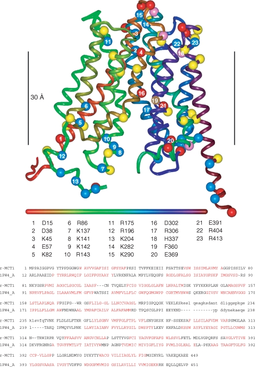
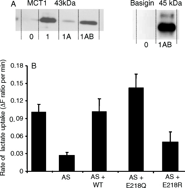
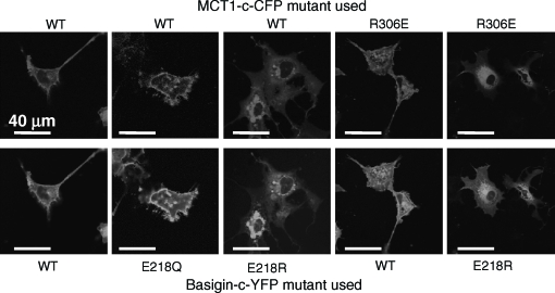
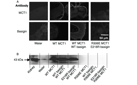
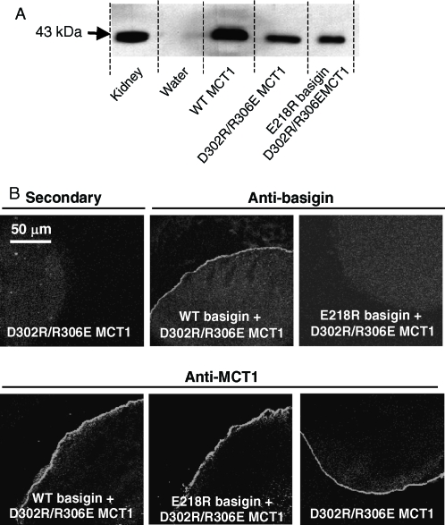
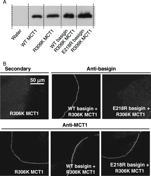
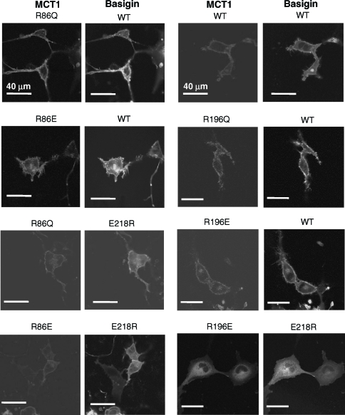
Monocarboxylate transporters MCT1-MCT4 require basigin (CD147) or embigin (gp70), ancillary proteins with a glutamate residue in their single transmembrane (TM) domain, for plasma membrane (PM) expression and activity. Here we use site-directed mutagenesis and expression in COS cells or Xenopus oocytes to investigate whether this glutamate (Glu218 in basigin) may charge-pair with a positively charged TM-residue of MCT1. Such residues were predicted using a new molecular model of MCT1 based upon the published structure of the E. coli glycerol-3-phosphate transporter. No evidence was obtained for Arg306 (TM 8) of MCT1 and Glu218 of basigin forming a charge-pair; indeed E218Q-basigin could replace WT-basigin, although E218R-basigin was inactive. No PM expression of R306E-MCT1 or D302R-MCT1 was observed but D302R/R306D-MCT1 reached the PM, as did R306K-MCT1. However, both were catalytically inactive suggesting that Arg306 and Asp302 form a charge-pair in either orientation, but their precise geometry is essential for catalytic activity. Mutation of Arg86 to Glu or Gln within TM3 of MCT1 had no effect on plasma membrane expression or activity of MCT1. However, unlike WT-MCT1, these mutants enabled expression of E218R-basigin at the plasma membrane of COS cells. We propose that TM3 of MCT1 lies alongside the TM of basigin with Arg86 adjacent to Glu218 of basigin. Only when both these residues are positively charged (E218R-basigin with WT-MCT1) is this interaction prevented; all other residue pairings at these positions may be accommodated by charge-pairing or stabilization of unionized residues through hydrogen bonding or local distortion of the helical structure.
Figures

Similar articles
-
Basigin (CD147) is the target for organomercurial inhibition of monocarboxylate transporter isoforms 1 and 4: the ancillary protein for the insensitive MCT2 is EMBIGIN (gp70).J Biol Chem. 2005 Jul 22;280(29):27213-21. doi: 10.1074/jbc.M411950200. Epub 2005 May 24. J Biol Chem. 2005. PMID: 15917240
-
Hydrophobic interactions stabilize the basigin-MCT1 complex.Protein J. 2009 Oct;28(7-8):362-8. doi: 10.1007/s10930-009-9202-3. Protein J. 2009. PMID: 19760495
-
Membrane-anchored carbonic anhydrase IV interacts with monocarboxylate transporters via their chaperones CD147 and GP70.J Biol Chem. 2019 Jan 11;294(2):593-607. doi: 10.1074/jbc.RA118.005536. Epub 2018 Nov 16. J Biol Chem. 2019. PMID: 30446621 Free PMC article.
-
The monocarboxylate transporter family--Structure and functional characterization.IUBMB Life. 2012 Jan;64(1):1-9. doi: 10.1002/iub.573. Epub 2011 Nov 30. IUBMB Life. 2012. PMID: 22131303 Review.
-
The SLC16 gene family - structure, role and regulation in health and disease.Mol Aspects Med. 2013 Apr-Jun;34(2-3):337-49. doi: 10.1016/j.mam.2012.05.003. Mol Aspects Med. 2013. PMID: 23506875 Review.
Cited by
-
Unassembled CD147 is an endogenous endoplasmic reticulum-associated degradation substrate.Mol Biol Cell. 2012 Dec;23(24):4668-78. doi: 10.1091/mbc.E12-06-0428. Epub 2012 Oct 24. Mol Biol Cell. 2012. PMID: 23097496 Free PMC article.
-
Monocarboxylate transporters in cancer.Mol Metab. 2020 Mar;33:48-66. doi: 10.1016/j.molmet.2019.07.006. Epub 2019 Jul 27. Mol Metab. 2020. PMID: 31395464 Free PMC article. Review.
-
Basigin Associates with Integrin in Order to Regulate Perineurial Glia and Drosophila Nervous System Morphology.J Neurosci. 2020 Apr 22;40(17):3360-3373. doi: 10.1523/JNEUROSCI.1397-19.2020. Epub 2020 Apr 7. J Neurosci. 2020. PMID: 32265259 Free PMC article.
-
Insights into molecular properties of the human monocarboxylate transporter 8 by combining functional with structural information.Thyroid Res. 2011 Aug 3;4 Suppl 1(Suppl 1):S4. doi: 10.1186/1756-6614-4-S1-S4. Thyroid Res. 2011. PMID: 21835051 Free PMC article.
-
Overview of the proton-coupled MCT (SLC16A) family of transporters: characterization, function and role in the transport of the drug of abuse gamma-hydroxybutyric acid.AAPS J. 2008 Jun;10(2):311-21. doi: 10.1208/s12248-008-9035-6. Epub 2008 Jun 4. AAPS J. 2008. PMID: 18523892 Free PMC article. Review.
References
-
- Abramson J, Smirnova I, Kasho V, Verner G, Kaback HR, Iwata S. Structure and mechanism of the lactose permease of Escherichia coli. Science. 2003;301:610–615. - PubMed
-
- Donovan JA, Jennings ML. N-Hydroxysulphosuccinimido active esters and the L-(+)-lactate transport protein in rabbit erythrocytes. Biochemistry. 1986;25:1538–1545. - PubMed
-
- Finnemann SC, Marmorstein AD, Neill JM, Rodriguez-Boulan E. Identification of the retinal pigment epithelium protein RET-PE2 as CE-/OX-47, a member of the immunoglobulin superfamily. Invest Ophthalmol Vis Sci. 1997;38:2366–2374. - PubMed
-
- Friesema ECH, Ganguly S, Abdalla A, Fox JEM, Halestrap AP, Visser TJ. Identification of monocarboxylate transporter 8 as a specific thyroid hormone transporter. J Biol Chem. 2003;278:40128–40135. - PubMed
Publication types
MeSH terms
Substances
Grants and funding
LinkOut - more resources
Full Text Sources
Molecular Biology Databases