. 2006 Dec 5;45(48):14379-88.
doi: 10.1021/bi0617355.

Specific effects of potassium ion binding on wild-type and L358P cytochrome P450cam

Affiliations

Specific effects of potassium ion binding on wild-type and L358P cytochrome P450cam

Bo OuYang et al. Biochemistry. .

Abstract

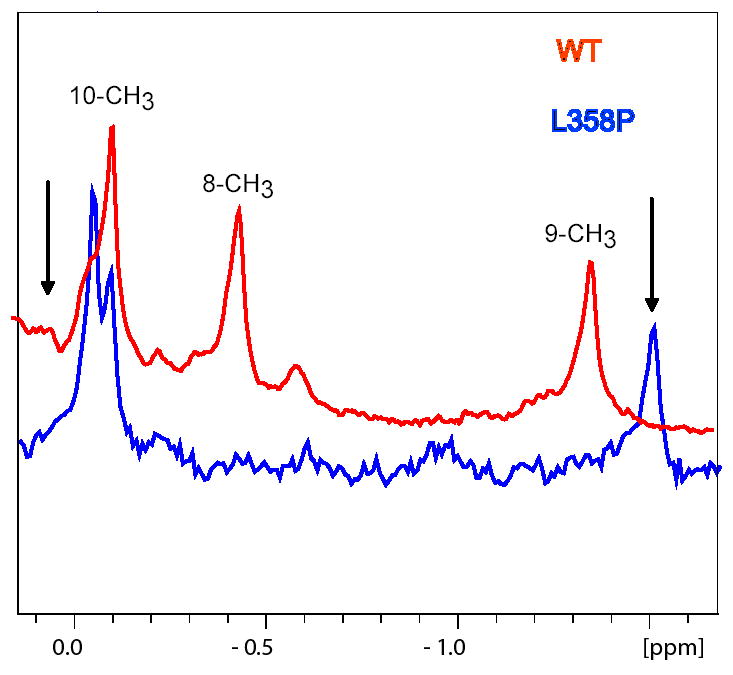
The camphor monoxygenase cytochrome P450cam (CYP101) requires potassium ion (K+) to drive formation of the characteristic high-spin state of the heme Fe+3 upon substrate binding. Amide 1H, 15N correlations in perdeuterated [U-15N] CYP101 were monitored as a function of K+ concentration by 2D-TROSY-HSQC in both camphor-bound oxidized (CYP-S) and camphor- and CO-bound reduced CYP101 (CYP-S-CO). In both forms, K+-induced spectral perturbations are detected in the vicinity of the K+ binding site proposed from crystallographic structures, but are larger and more widespread structurally in CYP-S than in CYP-S-CO. In CYP-S-CO, K+-induced perturbations occur primarily near the proposed K+ binding site in the B-B' loop and B' helix, which are also perturbed by binding of effector, putidaredoxin (Pdx). The spectral effects of K+ binding in CYP-S-CO oppose those observed upon Pdxr titration. However, Pdxr titration of CYP-S-CO in the absence of K+ results in multiple conformations. The spin-state equilibrium in the L358P mutant of CYP101 is more sensitive to K+ concentration than WT CYP101, consistent with a hypothesis that L358P preferentially populates conformations enforced by Pdx binding in WT CYP101. Thallium(I), a K+ mimic, minimizes the effects of Pdx titration on the NMR spectrum of CYP-S-CO, but is competent to replace K+ in driving the formation of high-spin CYP-S. These observations suggest that the role of K+ is to stabilize conformers of CYP-S that drive the spin-state change prior to the first electron transfer, and that K+ stabilizes the CYP-S-CO conformer that interacts with Pdx. However, upon binding of Pdx, further conformational changes occur that disfavor K+ binding.


Figures

Figure 1
Figure 1
Left) Second-derivative UV-visible spectra of oxidized WT CYP-S showing the effect of K+ concentration on spin state equilibrium. Black curve was obtained at 0 mM KCl, red curve at 50 mM KCl and blue curve in 100 mM KCl. All spectra were recorded at 25°C with 6 μM CYP101 in 50 mM Tris-HCl, 1 mM camphor, pH 7.4. Right) Second-derivative UV-visible spectra of oxidized L358P CYP-S. Black curve was obtained at 0 mM KCl, red curve at 50 mM KCl and blue curve in 100 mM KCl. Spectra were recorded at 25°C with 3 μM L358P CYP101 in 50 mM Tris-HCl, 1 mM camphor, pH 7.4.
Figure 2
Figure 2
Comparison of K+ and Na+ salt effects on 1H, 15N correlations in TROSY-HSQC spectra of reduced CYP-S-CO (upper spectra) and oxidized CYP-S (lower spectra). Spectra on the left show the amide NH correlation of Cys 85, adjacent to carbonyl of Glu 84, proposed to ligate K+ (). Upper left is reduced, lower left is oxidized. No correlation for Cys 85 is observed in the absence of alkali chloride salt in CYP-S (lower left). Spectra on the right show differential effects in reduced CYP-S-CO and oxidized CYP-S for Gly 226, located in the β2 sheet. All spectra were obtained at 25 °C, 600 MHz in 50 mM Tris-HCl, 2 mM camphor, pH 7.4.
Figure 3
Figure 3
NMR spectral perturbations induced by K+ titration in CYP-S-CO and CYP-S mapped onto the CYP101 structure. Both figures are based on the 3CPP structure of Raag and Poulos (). WAT 515, presumed to be K+ in the 3CPP structure, is indicated by a red sphere in both structures. Heme and camphor are in salmon. Left) Reduced CYP-S-CO. Backbone positions marked in red are perturbed and include the B-B’ loop and B’ helix including K+ binding residues Glu 84, Gly 93, Glu 94 and Tyr 96. Those in dark blue are perturbed by >10 Hz in either 15N or 1H, and include portions of the β1 and β5 strands. See Table 2 for details. Right) Oxidized CYP-S. Backbone positions marked in red indicate residues that are either doubled or missing in the absence of K+. These include portions of the β1, β4 and β5 strands, the B-B’ loop, B’ helix, C helix, E helix, F-G loop and G helix. See Table 2 for details. Residues in white indicate paramagnetic bleaching in the oxidized CYP-S for which no data is available. Figures were generated using MOLSCRIPT ().
Figure 4
Figure 4
Comparison of the effects of K+ and Pdxr binding in the presence of K+ to 0.3 mM CYP-S-CO (90/10 H2O/D2O, 50 mM Tris-HCl pH 7.4, 2 mM camphor, 25 °C) on 1H,15N TROSY-HSCQ correlations obtained at 600 MHz 1H. Spectra shown are typical. Titration of K+-bound CYP-S-CO with Pdxr moves correlations affected by K+ binding in the direction of the K+-free form in both 15N and 1H dimensions.
Figure 5
Figure 5
Evidence for two conformers at slow exchange in CYP-S-CO in the presence of Pdxr and the absence of K+. Addition of 1 eq. of Pdxr to 0.5 mM K+-free CYP-S-CO (90/10 H2O/D2O, 50 mM Tris-HCl pH 7.4, 2 mM camphor, 25 °C) splits the NH correlations of Glu 40 and Glu 91 in 1H,15N TROSY-HSCQ correlations obtained at 600 MHz 1H. For comparison, the addition of 1 eq. of Pdxr to 0.3 mM CYP-S-CO in the presence of K+ is shown for Glu 91 in the bottom panel (90/10 H2O/D2O, 100 mM KCl, 50 mM Tris-HCl pH 7.4, 2 mM camphor, 25 °C).
Figure 6
Figure 6
Upfield regions of the 600 MHz 1H NMR spectra of perdeuterated WT (red) and L358P (blue) CYP-S-CO showing the three methyl resonances of enzyme-bound camphor (). Black arrows indicate the position of the 8-methyl (left, downfield) and 9-methyl (right, upfield) resonances in WT CYP-S-CO in the presence of 3 eq. of Pdxr. Sample conditions are 90/10 H2O/D2O, 50 mM Tris-HCl pH 7.4, 2 mM camphor, 25 °C.

Similar articles

Cited by

References

    1. Britt BM. For enzymes, bigger is better. Biophys Chem. 1997;69:63–70. - PubMed
    1. Mueller EJ, Loida PJ, Sligar SG. Cytochrome P450: Structure, Function and Biochemistry. 1995. pp. 83–124.
    1. Lange R, Bonfils C, Debey P. The low-spin reversible highspin transition equilibrium of camphor-bound cytochrome P-450. Effects of medium and temperature on equilibrium data. Eur J Biochem. 1977;79:623–628. - PubMed
    1. Lange R, Pierre J, Debey P. Visible and ultraviolet spectral transitions of camphor-bound cytochrome P-450. A comprehensive study. Eur J Biochem. 1980;107:441–445. - PubMed
    1. Hui Bon Hoa G, Marden MC. The pressure dependence of the spin equilibrium of camphor-bound cytochrome P450. Eur J Biochem. 1982;124:311–315. - PubMed

Publication types