. 2006 Dec 5;45(48):14533-42.
doi: 10.1021/bi0616652.

The N-terminal domain of Bcl-xL reversibly binds membranes in a pH-dependent manner

Affiliations

The N-terminal domain of Bcl-xL reversibly binds membranes in a pH-dependent manner

Guruvasuthevan R Thuduppathy et al. Biochemistry. .

Abstract

Bcl-xL regulates apoptosis by maintaining the integrity of the mitochondrial outer membrane by adopting both soluble and membrane-associated forms. The membrane-associated conformation does not require a conserved, C-terminal transmembrane domain and appears to be inserted into the bilayer of synthetic membranes as assessed by membrane permeabilization and critical surface pressure measurements. Membrane association is reversible and is regulated by the cooperative binding of approximately two protons to the protein. Two acidic residues, Glu153 and Asp156, that lie in a conserved hairpin of Bcl-xLDeltaTM appear to be important in this process on the basis of a 16% increase in the level of membrane association of the double mutant E153Q/D156N. Contrary to that for the wild type, membrane permeabilization for the mutant is not correlated with membrane association. Monolayer surface pressure measurements suggest that this effect is primarily due to less membrane penetration. These results suggest that E153 and D156 are important for the Bcl-xLDeltaTM conformational change and that membrane binding can be distinct from membrane permeabilization. Taken together, these studies support a model in which Bcl-xL activity is controlled by reversible insertion of its N-terminal domain into the mitochondrial outer membrane. Future studies with Bcl-xL mutants such as E153Q/D156N should allow determination of the relative contributions of membrane binding, insertion, and permeabilization to the regulation of apoptosis.


Figures

Figure 1
Figure 1
Bcl-xLΔTM associates and dissociates with lipid vesicles as a function of pH. The fraction of protein bound to lipid vesicles (Fbound) was measured by a vesicle sedimentation assay, and at pH 7.0, less than 10% of the Bcl-xLΔTM is membrane-associated. By contrast, when this sample is titrated to pH 4.0 with concentrated HCl, more than 95% of the Bcl-xLΔTM is membrane-associated. When this sample is titrated back to pH 7.0 with concentrated NaOH, less than 5% of the Bcl-xLΔTM is membrane-associated, suggesting that Bcl-xLΔTM reversibly associates with lipid vesicles. The data are means ± the standard error of the mean of at least three independent measurements using different lipid vesicle and protein preparations.
Figure 2
Figure 2
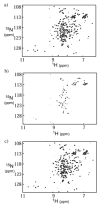
Solution conformation of Bcl-xLΔTM which is unaltered by membrane association as assessed by two-dimensional NMR spectroscopy. (a) 15N-labeled Bcl-xLΔTM (60 μM) was incubated overnight with lipid vesicles (60:40 DOPC:DOPG ratio at 25 °C and pH 7.25) at a protein:lipid ratio of 1:200 before a 1H–15N HSQC spectrum was recorded at 14.1 T with a cryoprobe. (b) After data collection, the sample was titrated to pH 4.65 by adding small amounts of concentrated HCl and incubated for 10 min before another HSQC spectrum was recorded. Under these conditions, the majority of NMR signals disappear presumably due to the increased rate of transverse relaxation for the protein associated with the lipid vesicle. (c) This sample was then titrated to pH 7.4 by the addition of small amounts of concentrated NaOH and incubated for 10 min. The subsequent HSQC spectrum exhibits the return of the NMR signals with chemical shifts that appear similar to those of the spectrum before acidification.
Figure 3
Figure 3
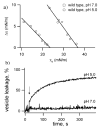
Bcl-xLΔTM increases the surface pressure of lipid monolayers and permeabilizes lipid vesicles in a pH-dependent manner. (a) Changes in surface pressure (Δ π) of lipid monolayers (60:40 DOPC:DOPG) after addition of Bcl-xLΔTM to the subphase are measured as function of the initial surface pressure (πo) at pH 7.0 (□) and 5.0 (○). The data are fit to a straight line, and the x-intercepts correspond to the monolayer critical surface pressure (πc), which provides a measure of the membrane penetrability of the protein (). For pH 7.0, the value of πc is 23.6 ± 1.2 mN/m, which increases to 36.9 ± 1.4 mN/m at pH 5.0. The uncertainty reflects the 95% confidence intervals in πc from a nonlinear regression analysis of the data. (b) The ability of Bcl-xLΔTM to cause membrane permeability was assessed at pH 7.0 and 5.0 by a dye leakage assay using ANTS and DPX encapsulated into lipid vesicles (12.5 and 45 mM, respectively, with a final lipid concentration of 0.1 mM) at 37 °C. At pH 7.0, little leakage of ANTS was detected after addition of Bcl-xLΔTM (denoted with an arrow). At pH 5.0, more than 60% of the ANTS was released after Bcl-xLΔTM addition. The kinetic traces are representative of at least three independent measurements using two different lipid vesicle preparations. The percentage of leakage was calculated after the complete release of the fluorescent probe by the addition of Triton X-100 [final concentration of 0.1% (w/v)].
Figure 4
Figure 4
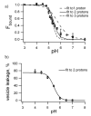
Cooperative pH dependence of the solution to membrane conformational change for Bcl-xLΔTM. (a) The level of association of protein with lipid vesicles (60:40 DOPC:DOPG) was measured as a function of pH by a sedimentation assay. The data are fit to a simplified thermodynamic linkage model that couples protonation to the conformational change. This simplified model assumes protein binds lipid vesicles only upon protonation by any number of protons, nH, that are governed by a single apparent pKaapp value. From the fits to the data, the number of protons bound to protein (nH) for the conformational change was estimated to be ~2 (—). For comparison, the fits to the data for an nH of 1 (– – –) and an nH of 3 (- - -) are shown. The values for Kx and pKaapp are interdependent and therefore cannot be determined accurately (see the text). (b) Cooperative pH dependence of Bcl-xLΔTM-induced leakage of lipid vesicles. The extent of leakage of the fluorescence dye ANTS from lipid vesicles (60:40 DOPC:DOPG) as a function of pH was measured as in Figure 4. The data are reasonably well fit to a similar linkage model with two protons (—).
Figure 5
Figure 5
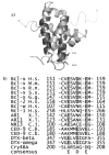
Conserved, hydrophobic helical hairpin of Bcl-xLΔTM that shares acidic residues with bacterial toxins and other Bcl-2 proteins. (a) The so-called hydrophobic, helical hairpin of Bcl-xLΔTM (α-helices 5 and 6 in darker gray) is postulated to mediate membrane association on the basis of considerations from the bacterial toxins. Residues E153 and D156 in the hairpin are highlighted using a stick representation. The protein is depicted without a large unstructured loop between residues D29 and R77, which is present in the protein used in our experiments. This figure was created using PYMOL (). (b) Sequence analysis of Bcl-2 proteins and bacterial toxins features acidic residues in the tip of the hairpin between hydrophobic α-helices.
Figure 6
Figure 6
E153Q/D156N mutant which differs from wild-type Bcl-xLΔTM in its pH dependence of membrane association and permeabilization. (a) Binding of protein to lipid vesicles as a function of pH was repeated for Bcl-xLΔTM (○) concurrently with measurements for E153Q/D156N (•) using the vesicle sedimentation assay as described in the legend of Figure 4a. (b) The difference between the binding of the mutant and wild type to lipid vesicles is shown for pH 4.0–7.0. At each pH that was measured, E153Q/D156N associated as well or better than wild-type Bcl-xLΔTM. This increase is roughly uniform at each pH between 5.0 and 7.0 (16 ± 3%). (c) The ability of the E153Q/D156N mutant to induce membrane permeabilization and its pH dependence was also tested. The extent of leakage of the fluorescence dye ANTS from lipid vesicles (60:40 DOPC:DOPG) as a function of pH was measured as described in the legend of Figure 4b. (d) The difference between the mutant and wild type inducing the release of ANTS from lipid vesicles is shown for pH 4.0–7.0. Above pH 5.5, E153Q/D156N induces more ANTS release from lipid vesicles than the wild type. However, below pH 5.5, E153Q/D156N induces less ANTS release from lipid vesicles than the wild type. Each datum is the mean ± the standard error of the mean of at least three independent measurements using two different lipid vesicle preparations.
Figure 7
Figure 7
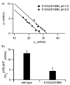
E153Q/D156N mutant which differs from wild-type Bcl-xLΔTM in the pH-dependent changes in monolayer surface pressure. (a) Changes in surface pressure (Δ π) of a lipid monolayer at pH 7.0 and 5.0 for E153Q/D156N were measured as described in the legend of Figure 3. At pH 7.0, critical surface pressure πc increased to 28.1 ± 1.2 mN/m for the mutant, while at pH 5.0, this value decreased to 32.5 ± 1.4 mN/m. (b) These differences are reflected in the pH-dependent change in the critical surface pressure for the lipid monolayer between pH 5.0 and 7.0 (Δ πcpH5–pH7), which decreases for the E153Q/D156N mutant by 3-fold compared to that of wild-type Bcl-xLΔTM. The error bars reflect the 95% confidence intervals in πc from a nonlinear regression analysis of the data.
None

Similar articles

Cited by

References

    1. Chao DT, Korsmeyer SJ. BCL-2 Family: Regulators of Cell Death. Annu Rev Immunol. 1998;16:395–419. - PubMed
    1. Gross A. BCL-2 Proteins: Regulators of the Mitochondrial Apoptotic Program. IUBMB Life. 2001;52:231–6. - PubMed
    1. Harris MH, Thompson CB. The Role of the Bcl-2 Family in the Regulation of Outer Mitochondrial Membrane Permeability. Cell Death Differ. 2000;7:1182–91. - PubMed
    1. Kuwana T, Newmeyer DD. Bcl-2-Family Proteins and the Role of Mitochondria in Apoptosis. Curr Opin Cell Biol. 2003;15:691–9. - PubMed
    1. Schendel SL, Montal M, Reed JC. Bcl-2 Family Proteins as Ion Channels. Cell Death Differ. 1998;5:372–80. - PubMed

Publication types