. 2007 Apr 1;403(1):89-95.
doi: 10.1042/BJ20061738.

Lysosomes and Fas-mediated liver cell death

Affiliations

Lysosomes and Fas-mediated liver cell death

Robert Wattiaux et al. Biochem J. .

Abstract

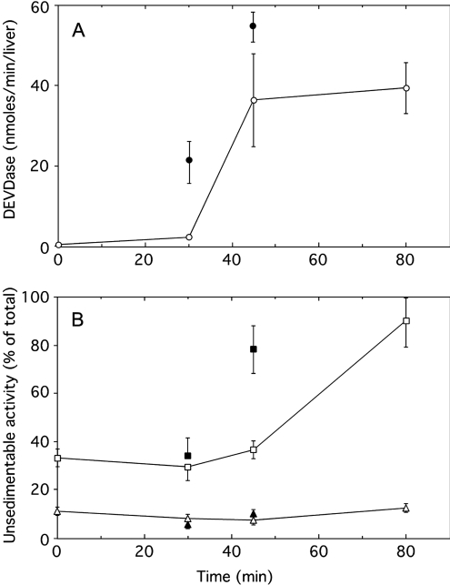
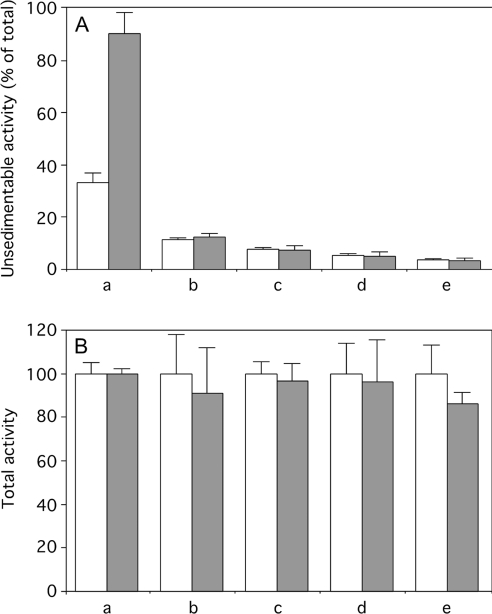
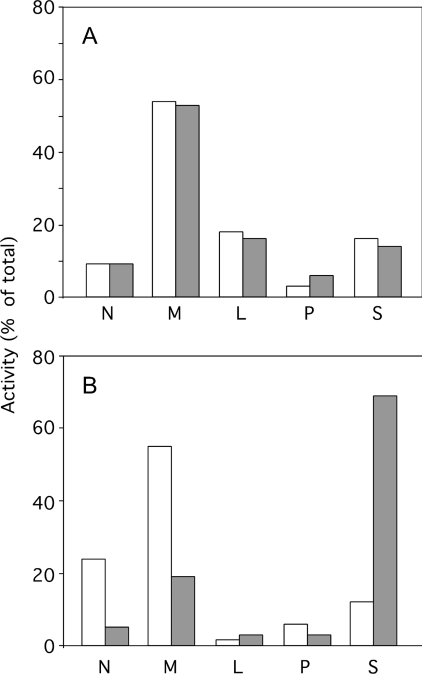
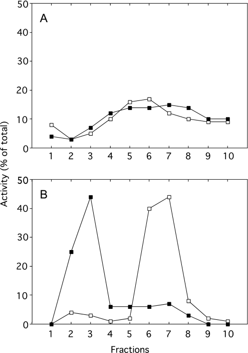
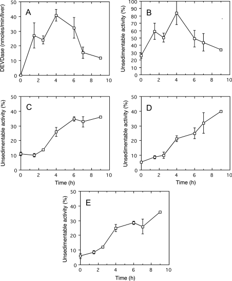
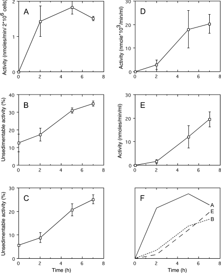
A number of studies, mostly performed ex vivo, suggest that lysosomes are involved in apoptosis as a result of a release of their cathepsins into the cytosol. These enzymes could then contribute to the permeabilization of the outer mitochondrial membrane; they could also activate effector caspases. The present study aims at testing whether the membrane of liver lysosomes is disrupted during Fas-mediated cell death of hepatocytes in vivo, a process implicated in several liver pathologies. Apoptosis was induced by injecting mice with aFas (anti-Fas antibody). The state of lysosomes was assessed by determining the proportion of lysosomal enzymes (beta-galactosidase, beta-glucuronidase, cathepsin C and cathepsin B) present in homogenate supernatants, devoid of intact lysosomes, and by analysing the behaviour in differential and isopycnic centrifugation of beta-galactosidase. Apoptosis was monitored by measuring caspase 3 activity (DEVDase) and the release of sulfite cytochrome c reductase, an enzyme located in the mitochondrial intermembrane space. Results show that an injection of 10 microg of aFas causes a rapid and large increase in DEVDase activity and in unsedimentable sulfite cytochrome c reductase. This modifies neither the proportion of unsedimentable lysosomal enzyme in the homogenates nor the behaviour of lysosomes in centrifugation. Experiments performed with a lower dose of aFas (5 microg) indicate that unsedimentable lysosomal hydrolase activity increases in the homogenate after injection but with a marked delay with respect to the increase in DEVDase activity and in unsedimentable sulfite cytochrome c reductase. Comparative experiments ex vivo performed with Jurkat cells show an increase in unsedimentable lysosomal hydrolases, but much later than caspase 3 activation, and a release of dipeptidyl peptidase III and DEVDase into culture medium. It is proposed that the weakening of lysosomes observed after aFas treatment in vivo and ex vivo results from a necrotic process that takes place late after initiation of apoptosis.


Figures

Figure 1
Figure 1. Caspase-3-like protease (DEVDase) activation (A) and unsedimentable activities (B) of sulfite cytochrome c reductase (□) and β-galactosidase (△) at increasing time after injection of aFas (10 μg/animal)
Determinations were performed on total homogenate for DEVDase, on total homogenate and on high-speed supernatant (35000 rev./min for 40 min; Beckman rotor type 40) for sulfite cytochrome c reductase and β-galactosidase. DEVDase activity was given in nmol of methylumbelliferone released/min per liver. The unsedimentable activity was expressed as percentage of the total activity present in the homogenate. Error bars represent the S.E.M. for at least three animals. Closed symbols give the values obtained after stimulation of apoptosis by an intraperitoneal injection of fructose (250 mg) 15 min before aFas injection.
Figure 2
Figure 2. Unsedimentable (A) and total (B) activities of sulfite cytochrome c reductase (a), β-galactosidase (b), β-glucuronidase (c), cathepsin C (d) and cathepsin B (e) present in liver homogenates originating from mice injected with saline (open bars) or with aFas (10 μg/animal) (closed bars) and killed 80 min after injection
Unsedimentable activity was expressed as a percentage of the activity present in the homogenate. For sulfite cytochrome c reductase and β-galactosidase, values indicated in Figure 1 have been used. Values for unsedimentable activity of sulfite cytochrome c reductase of treated versus control mice are significantly different (P<0.01) but not for unsedimentable lysosomal hydrolases (P>0.05). To make the presentation uniform, total activity measured in the aFas-injected mice liver homogenates is given as a percentage of the activity found in control mice liver homogenates supposed to be equal to 100. Error bars represent the S.E.M. for at least three animals. Values found in homogenates of injected and control mice do not significantly differ (P>0.05).
Figure 3
Figure 3. Distribution of β-galactosidase (A) and sulfite cytochrome c reductase (B) after differential centrifugation in 0.25 M sucrose
N, nuclear fraction; M, heavy mitochondrial fraction; L, light mitochondrial fraction; P, microsomal fraction; S, soluble fraction. Liver originated from a saline-injected mouse (open bars) or from an aFas-injected mouse killed 60 min after injection (closed bars). DEVDase activities of homogenates were respectively 0.1 nmol/min per liver for the control and 90.1 nmol/min per liver for the aFas-injected mouse.
Figure 4
Figure 4. Distribution of β-galactosidase (A) and cytochrome oxidase (B) after isopycnic centrifugation in a Percoll gradient, with 0.25 M sucrose as a solvent, of a total mitochondrial fraction (M+L) isolated from the liver of a saline-injected mouse (□) or of an aFas-injected mouse killed 60 min after injection (■)
The M+L fraction was layered at the top of the gradient. Centrifugation was performed at 25000 rev./min for 60 min in the Beckman rotor SW 65. Fractions are numbered from the top to the bottom of the gradient. DEVDase activities of homogenates were respectively 0 nmol/min per liver for the control and 0 and 66.1 nmol/min per liver for the aFas-injected mouse.
Figure 5
Figure 5. Caspase-3-like protease (DEVDase) activation (A) and unsedimentable activities of sulfite cytochrome c reductase (B), β-galactosidase (C), cathepsin C (D) and β-glucuronidase (E) at increasing time after injection of aFas (5 μg/animal)
Determinations were performed on total homogenate for DEVDase, on total homogenate and on high-speed supernatant (35000 rev./min for 40 min; Beckman rotor type 40) for sulfite cytochrome c reductase and acid hydrolases. DEVDase activity was given in nmol of methylumbelliferone released/min per liver. The unsedimentable activity was expressed as a percentage of the total activity present in the homogenate. Error bars represent the S.E.M. for at least three animals. The last values (9 h) were obtained in a single experiment.
Figure 6
Figure 6. Caspase-3-like protease (DEVDase) activation in cells (A), unsedimentable activities of β-galactosidase (B) and of cathepsin C (C) in cell homogenates, activities of DPPIII (D) and of DEVDase (E) in culture medium, at increasing times after addition of aFas (50 ng/ml) to Jurkat cell culture medium.
Determinations were performed on total homogenate of Jurkat cells and on the culture medium for DEVDase, on total homogenate and on high-speed supernatant for β-galactosidase and cathepsin C. DEVDase activity was given in nmol of methylumbelliferone released·min−1·(2×106 cells)−1 or ml of culture medium. DPPIII activity is given in nmol of naphthylamine released·min−1·(ml of culture medium)−1. The unsedimentable activity was expressed as a percentage of the total activity present in the homogenate. Error bars represent the S.E.M. for five cell preparations. (F) Outline of (A, B, E) to compare the time courses of apoptosis (A), lysosome leakage (B) and cell lysis (E).

Similar articles

Cited by

References

    1. Guicciardi M. E., Leist M., Gores G. J. Lysosomes in cell death. Oncogene. 2004;23:2881–2890. - PubMed
    1. Cirma T., Oresi K., Mazovec G. D., Turk V., Reed J. C., Myers R. M., Salvesen G. S., Turk B. Selective disruption of lysosomes in Hela cells triggers apoptosis mediated by cleavage of Bid by multiple papain-like lysosomal cathepsins. J. Biol. Chem. 2003;279:3578–3587. - PubMed
    1. Ferri K. F., Kroemer G. Organelle-specific initiation of cell death pathways. Nat. Cell Biol. 2001;3:E255–E263. - PubMed
    1. Green D. R., Kroemer G. Pharmacological manipulation of cell death: clinical applications in sight? J. Clin. Invest. 2005;115:2610–2617. - PMC - PubMed
    1. Kroemer G., Martin S. J. Caspase-independent cell-death. Nat. Med. 2005;11:725–730. - PubMed

Publication types