. 2006 Nov 27:3:100.
doi: 10.1186/1743-422X-3-100.

Small interfering RNA targeted to stem-loop II of the 5' untranslated region effectively inhibits expression of six HCV genotypes

Affiliations

Small interfering RNA targeted to stem-loop II of the 5' untranslated region effectively inhibits expression of six HCV genotypes

Ramesh Prabhu et al. Virol J. .

Retraction in

Abstract

Background: The antiviral action of interferon alpha targets the 5' untranslated region (UTR) used by hepatitis C virus (HCV) to translate protein by an internal ribosome entry site (IRES) mechanism. Although this sequence is highly conserved among different clinical strains, approximately half of chronically infected hepatitis C patients do not respond to interferon therapy. Therefore, development of small interfering RNA (siRNA) targeted to the 5'UTR to inhibit IRES mediated translation may represent an alternative approach that could circumvent the problem of interferon resistance.

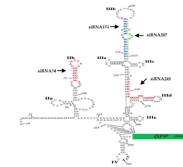
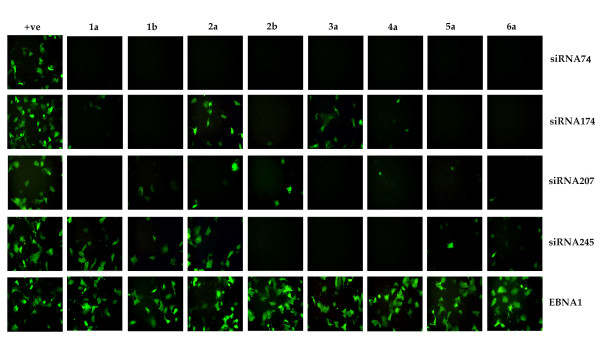
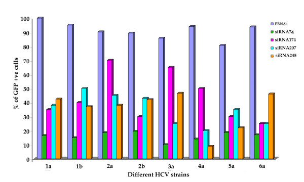
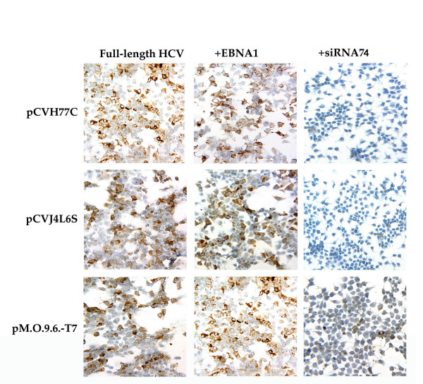
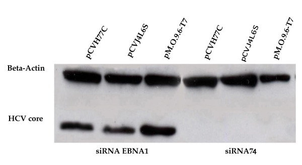
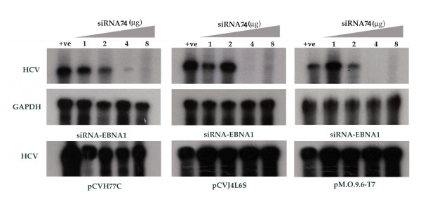
Results: Four different plasmid constructs were prepared for intracellular delivery of siRNAs targeting the stem loop II-III of HCV 5' UTR. The effect of siRNA production on IRES mediated translation was investigated using chimeric clones between the gene for green fluorescence protein (GFP) and IRES sequences of six different HCV genotypes. The siRNA targeted to stem loop II effectively mediated degradation of HCV IRES mRNA and inhibited GFP expression in the case of six different HCV genotypes, where as siRNAs targeted to stem loop III did not. Furthermore, intracytoplasmic expression of siRNA into transfected Huh-7 cells efficiently degraded HCV genomic RNA and inhibited core protein expression from infectious full-length infectious clones HCV 1a and HCV 1b strains.

Conclusion: These in vitro studies suggest that siRNA targeted to stem-loop II is highly effective inhibiting IRES mediated translation of the major genotypes of HCV. Stem-loop II siRNA may be a good target for developing an intracellular immunization strategy based antiviral therapy to inhibit hepatitis C virus strains that are not inhibited by interferon.


Figures

Figure 1
Figure 1
Location of siRNA targets to the 5'UTR of HCV genome. Predicted secondary structure of the 5' UTR sequences (18–357). The sequence shown is that of the genotype 1a 5'UTR, HCV-H [40], and the structure based on previous studies [41-43]. Stem-loop structures are labeled for reference. The chimeric clones were made by fusing the GFP-encoding sequence, including a poly (A) tail, after the CCU sequence of the 5'UTR by overlapping PCR. The locations of siRNA targets in the stem-loop regions are shown by arrows.
Figure 2
Figure 2
Effect of small interfering RNAs on the expression of green fluorescent protein from IRES clones of six different HCV genotypes. Huh-7 cells were co-transfected with HCVIRES-GFP plasmid with siRNA plasmid (pSuper-retro) by using the FuGENE 6 transfection reagent. After 48 hours transfected cells were observed under a fluorescence microscope. The experiments were repeated for each genotype IRES clones using different siRNA constructs. SiRNA-74 was the most effective and inhibited the expression of green fluorescence in all genotypes of HCV compared to other siRNA. Control siRNA targeted to EBNA1 had no effect on GFP expression from HCV IRES clones.
Figure 3
Figure 3
Quantitative measurement of GFP positive Huh-7 cells by flow cytometry after siRNA transfection. Huh-7 cells were co-transfected with HCVIRES-GFP plasmid with siRNA plasmid (pSuper-retro) by using the FuGENE 6 transfection reagent. After 48 hours of transfection, cells were harvested and GFP positive cells were analysed by a flow cytometer (Becton Dickinson, BD Biosciences, Clontech). Percentage of GFP-positive Huh-7 cells was quantitatively determined after siRNA transfection using cell quest computer software. The results were expressed as percentage of control. siRNA-74 was found to be most effective in silencing GFP from all IRES clones.
Figure 4
Figure 4
Immunocytochemical staining showing the silencing of core protein from full-length clones of HCV by siRNA-74. Huh-7 cells were co-transfected with (10 μg) pSuper-retro-siRNA74 and (10 μg) full-length clones of HCV. After 48 hours, transfected cells were harvested by the treatment with trypsin-EDTA. Cells were washed with PBS and immobilized onto a glass slide by cytospin method. Then slides were blocked and stained with core antibody against HCV using a mouse monoclonal antibody. Immunostaining for HCV core protein was performed using a standard protocol. The expression of core protein of full-length HCV 1a (pCVH77C), Ib (pCVJ4L6S), and 1b (pMO9.6--T7) is observed in the presence of siRNA74 and siRNAEBNA1 (unrelated siRNA).
Figure 5
Figure 5
Western blot analysis showing the silencing of core protein from full-length clones of HCV by siRNA transfection. Huh-7 cells were co-transfected with pSuper-retro-siRNA74 and full-length clones of HCV using the FuGENE 6 reagent. After 48 hours, transfected cells were isolated by the treatment with trypsin-EDTA. Cells were washed once with PBS and protein lysates were prepared and electrophoresed on 10% SDS-PAGE gels. The proteins were transferred to nitrocellulose membranes, blocked and immunoreacted with a primary antibody. The membrane was washed and incubated with peroxidase labeled secondary antibody and developed by ECL-chemiluminescence method. siRNA74 inhibited the expression of core protein in the case of all three full-length clones of HCV 1a and 1b. Beta actin levels were used as a control to make sure that equal amount of protein was present in the extracts.
Figure 6
Figure 6
Ribonuclease protection assay showing siRNA expression specifically degraded intracellular HCV positive strand RNA in the transfected Huh-7 cells. Huh-7 cells were co-transfected with different concentration of siRNA74 with different full-length clones of HCV. After 48 hours, transfected cells were isolated by the treatment with trypsin-EDTA. Total RNA was isolated and subjected to RPA for positive strand HCV using a minus strand RNA probe targeted to the 5'UTR region. The degradation of HCV positive strand by siRNA74 is concentration dependent. No HCV RNA degradation was observed in the cells transfected with unrelated siRNA.

Similar articles

Cited by

References

    1. Choo QL, Kuo G, Weiner AJ, Overby LR, Bradley DW, Houghton M. Isolation of a cDNA clone derived from a blood-borne non-A, non-B viral hepatitis genome. Science. 1989;244:359–362. doi: 10.1126/science.2523562. - DOI - PubMed
    1. Alter MJ. Epidemiology of hepatitis C. Hepatology. 1997;26:62S–65S. doi: 10.1002/hep.510260711. - DOI - PubMed
    1. World health Organization Hepatitis C: global prevalence (update) Wkly Epidemiol Rec. 2000;75:18–19. - PubMed
    1. Theodore D, Fried MW. Natural history and disease manifestations. Curr Top Microbiol. 2000;242:44–54. - PubMed
    1. Alter HJ, Seeff LB. Recovery, persistence and sequelae in hepatitis C virus infection: a perspective on long-term outcome. Seminar Liver Disease. 2000;20:17–35. doi: 10.1055/s-2000-9505. - DOI - PubMed

Publication types

LinkOut - more resources