. 2006;34(22):6461-71.
doi: 10.1093/nar/gkl820. Epub 2006 Nov 27.

Self-splicing of a group IIC intron: 5' exon recognition and alternative 5' splicing events implicate the stem-loop motif of a transcriptional terminator

Affiliations

Self-splicing of a group IIC intron: 5' exon recognition and alternative 5' splicing events implicate the stem-loop motif of a transcriptional terminator

Navtej Toor et al. Nucleic Acids Res. 2006.

Abstract

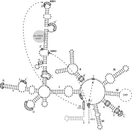
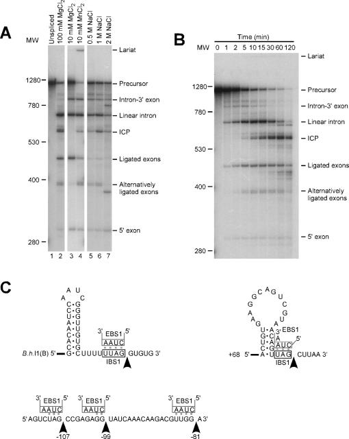
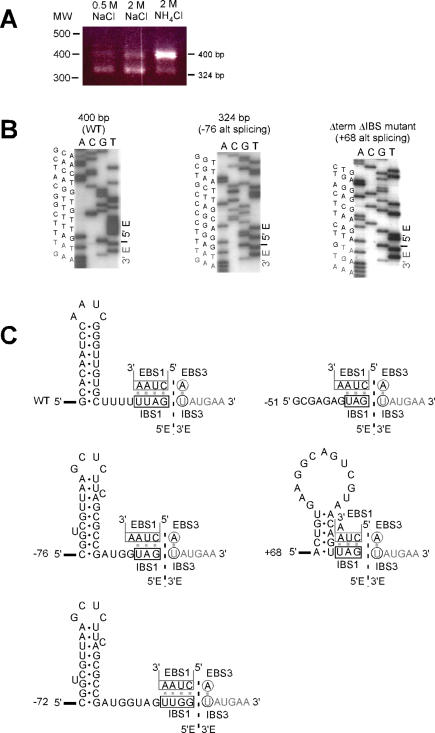
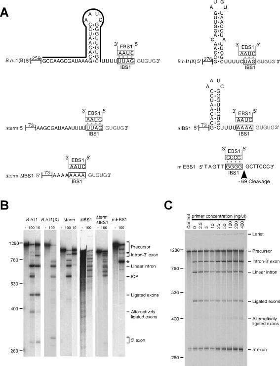
Bacterial IIC introns are a newly recognized subclass of group II introns whose ribozyme properties have not been characterized in detail. IIC introns are typically located downstream of transcriptional terminator motifs (inverted repeat followed by T's) or other inverted repeats in bacterial genomes. Here we have characterized the self-splicing activity of a IIC intron, B.h.I1, from Bacillus halodurans. B.h.I1 self-splices in vitro through hydrolysis to produce linear intron, but interestingly, additional unexpected products were formed that were highly dependent on ionic conditions. These products were determined to represent alternative splicing events at the 5' junction and cleavages throughout the RNA transcript. The alternative splicing and cleavage events occurred at cryptic splice sites containing stem-loop and IBS1 motifs, suggesting that the 5' exon is recognized by both elements. These results provide the first example of a group II intron that uses 5' splice sites nonadjacent to the ribozyme structure. Furthermore, the data suggest that IIC introns differ from IIA and IIB introns with respect to 5' exon definition, and that the terminator stem-loop substitutes in part for the missing IBS2-EBS2 (intron and exon binding sites 2) interaction.


Figures

Figure 1
Figure 1
Secondary structure of the B.h.I1(B) intron, a representative of group IIC introns. The six domains of the intron RNA structure are shown with black lettering, and exons with gray lettering. Domain IV encodes a 1.3 kb ORF, which is denoted by a dotted line. Tertiary interactions within the intron structure are indicated with Greek letters. Of these, the non-Watson–Crick interactions κ, θ, λ and ζ are predicted by analogy with other introns, but have not been confirmed for any IIC introns. The pairing interactions IBS1–EBS1, IBS3–EBS3 and γ–γ′ are involved in 5′ and 3′ exon recognition, and are highlighted by dotted lines. Like other IIC introns, B.h.I1(B) lacks an EBS2 motif in domain ID (gray circle), and the 5′ exon contains a terminator stem–loop in the position where the IBS2 is located in IIA and IIB introns.
Figure 2
Figure 2
Self-splicing products under different conditions and identification of bands. (A) B.h.I1(B) transcript was self-spliced in standard conditions of 100 mM MgCl2, 50 mM Tris (pH 7.5) and 2 M NH4Cl at 55°C for 15 min, or in alternative conditions with substitutions of 10 mM MgCl2 or 10 mM MnCl2 for 100 mM MgCl2, or 0.5, 1.0 or 2 M NaCl for 2 M NH4Cl. RNA size markers are indicated to the left in nucleotides, and product identifications to the right are explained in the text. (B) Time course of the self-splicing reaction under standard conditions, illustrating the initial formation of linear intron and ligated exons, and subsequent accumulation of other products. (C) Sequences and structures of the RNA cleavages produced in the self-splicing reaction. Arrows indicate cleavage sites at: the exon–intron junction of B.h. I1(B); the ICP cleavages at −107, −99 and −81 relative to the 3′ end of the intron; and the ICP cleavage at +68 relative to the 5′ end of the intron. EBS1 pairings of the intron are shown above the exon sequences.
Figure 3
Figure 3
Cognate and alternative exon ligation events during in vitro splicing reactions. (A) RT–PCR of exon junctions produced in self-splicing reactions. Self-splicing was under standard conditions with 2 M NH4Cl, or with 0.5 or 2 M NaCl. RT–PCR detected two splicing products of 400 bp and 324 bp, whose proportions varied with ionic conditions. (B) Sequencing of ligated exon products. Sequences are shown for the exon-exon junctions of the 400 bp and 324 bp bands in Panel A, and also for the major ligation product formed for the ΔterminatorΔIBS1 mutant described in Figure 4. (C) Sequences and structures of ligated exons. Structures are shown for: the exon–exon products of a cognate splicing reaction (WT); the upstream alternative splicing events at −76, −72, and −51 (relative to the exon–intron junction); and the internal splicing event +68 (relative to the exon–intron junction). EBS1 and EBS3 interactions are drawn above the exon sequences. The +68 stem–loop is present in the native secondary structure, although recognition of the stem may require unpairing of α–α′.
Figure 4
Figure 4
Mutations and sequence variations in motifs involved in 5′ exon recognition. (A) Diagram of variations tested: B.h.I1(B), wild-type exons; B.h.I1(X), exons from the B.h.I1(X) site with a mispaired stem rather than a canonical rho-independent terminator motif; Δterm, deleted terminator stem–loop; ΔIBS1, mutated IBS1 motif; ΔtermΔIBS1, deleted terminator stem–loop and mutated IBS1 motif; mEBS1, mutated EBS1 motif in the intron. Numbers indicate the length of the 5′ leader (vector sequence and native exon combined). The line above the B.h.I1(B) sequence shows the positions corresponding to the complementary oligonucleotide added to the reaction in Panel C. The arrow for mEBS1 indicates the cleavage produced during the splicing reaction. (B) Self-splicing reactions of the variants tested. Each set of lanes shows unspliced transcript incubated without magnesium (left) and intron transcript spliced under standard conditions with either 10 mM or 100 mM MgCl2. The asterisk denotes the cleavage at −69 in the 5′ exon, which was determined by primer extension. Unligated 5′ exons cannot be seen in panel B for the constructs Δterm, ΔIBS1, ΔtermΔIBS1 and mEBS1 because the exons are too short, and unligated 3′ exons are too short to be seen for all constructs. (C) Self-splicing in the presence of an oligonucleotide that pairs with the terminator stem–loop. An oligonucleotide complementary to the terminator stem–loop of B.h.I1 was added to intron transcript during the denaturation/renaturation step, to disrupt the stem–loop structure (Panel A). The disruption resulted in an increase in unligated 5′ exon, and intron-3′ exon intermediates.

References

    1. Robart A.R., Zimmerly S. Group II intron retroelements: function and diversity. Cytogenet. Genome Res. 2005;110:589–597. - PubMed
    1. Lambowitz A.M., Zimmerly S. Mobile group II introns. Annu. Rev. Genet. 2004;38:1–35. - PubMed
    1. Toro N. Bacteria and archaea group II introns: additional mobile genetic elements in the environment. Environ. Microbiol. 2003;5:143–151. - PubMed
    1. Belfort M., Derbyshire V., Parker M.M., Cousineau B., Lambowitz A.M. Mobile introns: pathways and proteins. In: Craig N.L., Craigie R., Gellert M., Lambowitz A.M., editors. Mobile DNA II. Washington DC: ASM Press; 2002. pp. 761–783.
    1. Bonen L., Vogel J. The ins and outs of group II introns. Trends Genet. 2001;17:322–331. - PubMed

Publication types