. 2007 Feb;27(3):1146-57.
doi: 10.1128/MCB.01009-06. Epub 2006 Nov 27.

Inactivation of CUG-BP1/CELF1 causes growth, viability, and spermatogenesis defects in mice

Affiliations

Inactivation of CUG-BP1/CELF1 causes growth, viability, and spermatogenesis defects in mice

Chantal Kress et al. Mol Cell Biol. 2007 Feb.

Abstract

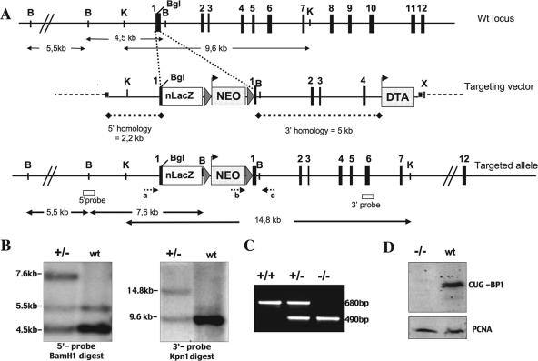
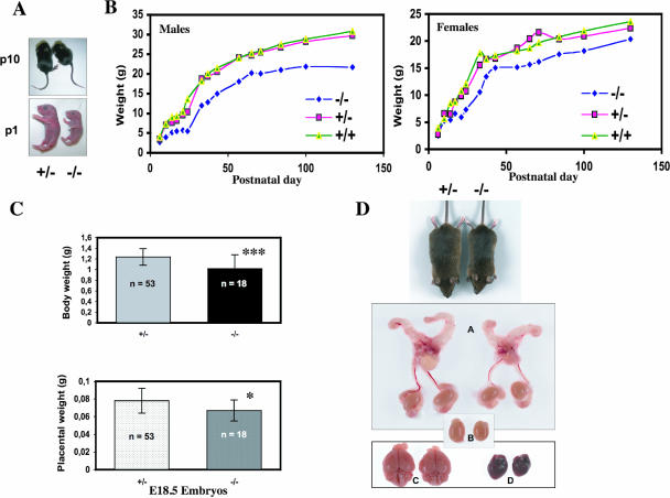
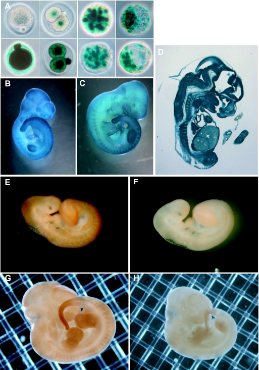
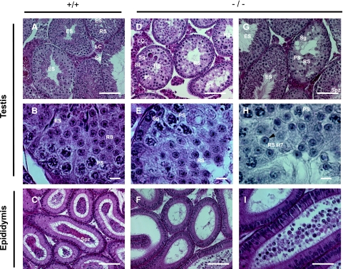
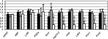
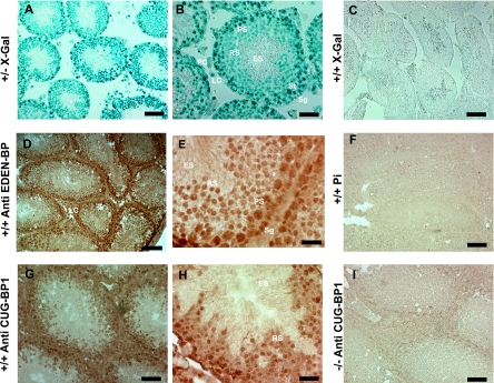
CUG-BP1/CELF1 is a multifunctional RNA-binding protein involved in the regulation of alternative splicing and translation. To elucidate its role in mammalian development, we produced mice in which the Cugbp1 gene was inactivated by homologous recombination. These Cugbp1(-/-) mice were viable, although a significant portion of them did not survive after the first few days of life. They displayed growth retardation, and most Cugbp1(-/-) males and females exhibited impaired fertility. Male infertility was more thoroughly investigated. Histological examination of testes from Cugbp1(-/-) males showed an arrest of spermatogenesis that occurred at step 7 of spermiogenesis, before spermatid elongation begins, and an increased apoptosis. A quantitative reverse transcriptase PCR analysis showed a decrease of all the germ cell markers tested but not of Sertoli and Leydig markers, suggesting a general decrease in germ cell number. In wild-type testes, CUG-BP1 is expressed in germ cells from spermatogonia to round spermatids and also in Sertoli and Leydig cells. These findings demonstrate that CUG-BP1 is required for completion of spermatogenesis.


Figures

FIG. 1.
FIG. 1.
Generation of Cugbp1-deficient mice. (A) Schematic representation of the genomic structure of the Cugbp1 locus (top), the targeting vector, and the targeted allele. Exons (solid boxes), LoxP sequences (triangles), and positions of primers for genotype analysis (small arrows a, b, and c) and probes used for Southern blots (open boxes) are indicated. The NEO box flanked with LoxP sequences and the DTA box represent positive and negative selection cassettes. Both are under a pgk promoter. The nls LacZ (nLacZ) cassette in frame with the Cugbp1 coding sequence downstream of the translation start site of Cugbp1 in exon 1 is also shown. Restriction sites relevant to the targeting construct and to the screening strategies are BamHI (B), KpnI (K), XhoI (X), and BglII (BgI). (B) Southern blot analysis of the recombinant and wild-type (wt) ES cell clones: genomic DNA digested with BamH1 or Kpn1 was subjected to Southern blotting and probed with the 5′ and 3′ probes, respectively, depicted in panel A. (C) Results of PCR genotyping using the primers (a, b, and c) shown in panel A. Amplification with primers a and c (wild-type allele) yielded a 680-bp PCR product, and amplification with primers a and c (recombinant allele) yielded a 490-bp PCR product. (D) Western blot of fibroblast whole-cell extract from mutant and wild-type mice, blotted with anti-CUG-BP1 monoclonal antibody (upper panel) or anti-PCNA antibody (lower panel).
FIG. 2.
FIG. 2.
Analysis of growth phenotype. (A) Representative pictures of heterozygous and Cugbp1/ littermates of newborn (P1) or 10-day-old (P10) mice. (B) Postnatal growth curve of male or female littermates. Mice were weighed over a period of 130 days. Results are plotted as the mean body weight of female −/− (n = 3 to 6), male −/− (n = 3 to 6), female +/− (n = 3 to 18), male +/− (n = 4 to 23), female +/+ (n = 3 to 6), or male +/+ (n = 3 to 10) mice. (C) Body or placenta weight of E18.5 +/− (n = 53) or −/− (n = 18) conceptuses. Fetal body weights (means ± standard deviation) were as follows: Cugbp1+/, 1.238 ± 0.153 g; Cugbp1/, 1.018 ± 0.259 g; ***, P < 0.001 (Fisher test). Placental weights (means ± standard deviations) were as follows: Cugbp1+/, 0,078 ± 0,014; Cugbp1/, 0,067 ± 0,012. *, P = 0.016 (Fisher test). (D) Macroscopic appearance of selected organs from a 4-month-old Cugbp1-deficient male (right) and a heterozygous male littermate (left). A, reproductive tract; B, testes; C, brain; D, heart.
FIG. 3.
FIG. 3.
CUG-BP1 expression during embryo development. (A to D) Expression of CUG-BP1 analyzed by X-Gal staining. (A) Upper panels, left, an oocyte from a +/+ female; then (left to right), two-cell, morulae, and blastocyst embryos derived from crosses between +/+ superovulated females and +− males. Lower panels, left, an oocyte from a +/− female; then (left to right), two-cell, morulae, and blastocyst embryos derived from reciprocal crosses between +/− superovulated females and +/+ males. (B) Ten-day-postcoitum embryo. (C) Eleven-day-postcoitum embryo. (D) Section of a 12-dpc embryo. (E to H) Expression of CUG-BP1 analyzed by immunohistochemistry of wild-type embryos. (E) Nine-day-postcoitum embryo stained with anti-EDEN-BP antibody. (F) Nine-day-postcoitum embryo stained with preimmune serum. (G) Twelve-day-postcoitum embryo stained with anti-EDEN-BP antibody. (H) Twelve-day-postcoitum embryo stained with preimmune serum.
FIG. 4.
FIG. 4.
Histological comparison of testes and epididymes from wild-type and Cugbp1/ mice. (A) Low-magnification section of a testis from a mature, wild-type male. (B) Higher magnification of the same section shown in panel A. (C) Low-magnification section of the epididymis from the same male. (D) Low-magnification section of a testis from a mature, Cugbp1/ male with a strong spermiogenesis arrest. (E) Higher magnification of the same section shown in panel D. (F) Low-magnification section of the epididymis from the same male. (G) Low-magnification section of a testis from a mature, Cugbp1/ male with a mild spermiogenesis arrest. (H) High-magnification section of a testis from a mature, Cugbp1/ male with a strong spermiogenesis arrest, stained with periodic acid Schiff and counterstained with hematoxylin. The arrowhead indicates the cap-shaped acrosome of the round spermatid (RS), and St7 is step 7 of spermiogenesis. (I) Higher magnification of F. Except for H, all the sections were stained with hematoxylin and eosin. LC, Leydig cells; Sg, spermatogonia; PS, pachytene spermatocyte; RS, round spermatid; ES, elongated spermatid; Spz, spermatozoa. For panels A, C, D, and F, bar = 100 μm; for panels B, E, and H, bar = 10 μm; for panels G and I, bar = 50 μm.
FIG. 5.
FIG. 5.
Apoptosis in testes of Cugbp1+/+ and Cugbp1/ males. Sections of testes from +/+ (A) or −/− (B) males were subjected to TUNEL staining. Spermatogonia (Sg) and pachytene spermatocyte (PS) and a round spermatid (RS) in B that are TUNEL positive are depicted. (C) Percentages of seminiferous tubules containing at least one TUNEL-positive cell in +/+ or −/− testes. P = 1.7 E−06, Student test. (D) Average number of TUNEL-positive cells per tubule containing at least one positive cell. P = 5 E−04, Student test.
FIG. 6.
FIG. 6.
Expression of Sertoli, Leydig, and germ cell markers. Relative quantities of the ubiquitously expressed GAPDH mRNA and of the indicated mRNAs were quantified by real-time RT-PCR. The amounts of the indicated mRNA relative to that of the GAPDH mRNA were calculated for +/+ (black bars; n = 3), +/− (gray bars; n = 4), and −/− (white bars; n = 3 [2 −/− mice with a histological “strong phenotype” and 1 mouse with a histological “mild phenotype”]; see Fig. 4) mice. Results are expressed as the mean (± standard deviation) of the ratios for each genotype, normalized to 1 for the average value of the three phenotypes. Statistical comparison between the +/+ genotype and the other genotypes was done with a Student test. When the P values were less than 0.1, they were indicated above the corresponding bars. ABP, androgen binding protein; LHR, LH receptor; PGDs, PGD synthetase; Acro, proacrosin; Prm1 and Prm2, protamines 1 and 2.
FIG. 7.
FIG. 7.
Expression of CUG-BP1 in the testis. (A and B) Testes from heterozygous males were used to analyze promoter activity of Cugbp1 by detection of β-galactosidase activity. B is a higher magnification of A. Localization of the staining is nuclear because of the presence of nls in the construction. (C) Testis from a wild-type (+/+) male was used as a negative control for X-Gal staining. (D to H) Testes from wild-type (+/+) mice were used for immunohistochemical localization of CUG-BP1. Immunohistochemistry was performed with an anti-EDEN-BP antibody (D and E), the corresponding preimmune serum (F), or anti-CUG-BP1 antibody (G and H). (I) Immunohistochemical staining performed on a testis from a −/− mouse with the anti-CUG-BP1 antibody. In panels D to G and I, testes were fixed in paraformaldehyde after congelation, whereas in panel H, the testis was fixed before congelation, which allowed preservation of the cytoplasmic compartment. LC, Leydig cell; St, Sertoli cell; PC, peritubular cell; Sg, spermatogonia; PS, pachytene spermatocyte; RS, round spermatid; ES, elongated spermatid. For panels A, D, C, and F, bar = 100 μm; for panels B, G, and I, bar = 50 μm; for panels E and H, bar = 25 μm.

Similar articles

Cited by

References

    1. Barreau, C., L. Paillard, A. Mereau, and H. B. Osborne. 2006. Mammalian CELF/Bruno-like RNA-binding proteins: molecular characteristics and biological functions. Biochimie 88:515-525. - PubMed
    1. Barreau, C., T. Watrin, H. Beverley Osborne, and L. Paillard. 2006. Protein expression is increased by a class III AU-rich element and tethered CUG-BP1. Biochem. Biophys. Res. Commun. 347:723-730. - PubMed
    1. Beck, A. R., I. J. Miller, P. Anderson, and M. Streuli. 1998. RNA-binding protein TIAR is essential for primordial germ cell development. Proc. Natl. Acad. Sci. USA 95:2331-2336. - PMC - PubMed
    1. Behr, R., and G. F. Weinbauer. 2001. cAMP response element modulator (CREM): an essential factor for spermatogenesis in primates? Int. J. Androl. 24:126-135. - PubMed
    1. Blendy, J. A., K. H. Kaestner, G. F. Weinbauer, E. Nieschlag, and G. Schutz. 1996. Severe impairment of spermatogenesis in mice lacking the CREM gene. Nature 380:162-165. - PubMed

Publication types

MeSH terms