

C57BL/6 and congenic interleukin-10-deficient mice can serve as models of Campylobacter jejuni colonization and enteritis
- PMID: 17130251
- PMCID: PMC1828563
- DOI: 10.1128/IAI.00833-06
C57BL/6 and congenic interleukin-10-deficient mice can serve as models of Campylobacter jejuni colonization and enteritis
Abstract
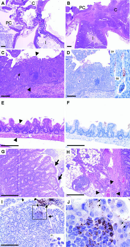
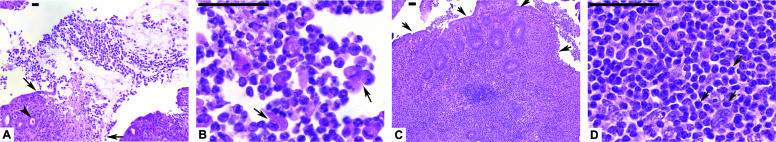
Campylobacter jejuni is a globally distributed cause of human food-borne enteritis and has been linked to chronic joint and neurological diseases. We hypothesized that C. jejuni 11168 colonizes the gastrointestinal tract of both C57BL/6 mice and congenic C57BL/6 interleukin-10-deficient (IL-10(-/-)) mice and that C57BL/6 IL-10(-/-) mice experience C. jejuni 11168-mediated clinical signs and pathology. Individually housed mice were challenged orally with C. jejuni 11168, and the course of infection was monitored by clinical examination, bacterial culture, C. jejuni-specific PCR, gross pathology, histopathology, immunohistochemistry, and anti-C. jejuni-specific serology. Ceca of C. jejuni 11168-infected mice were colonized at high rates: ceca of 50/50 wild-type mice and 168/170 IL-10(-/-) mice were colonized. In a range from 2 to 35 days after infection with C. jejuni 11168, C57BL/6 IL-10(-/-) mice developed severe typhlocolitis best evaluated at the ileocecocolic junction. Rates of colonization and enteritis did not differ between male and female mice. A dose-response experiment showed that as little as 10(6) CFU produced significant disease and pathological lesions similar to responses seen in humans. Immunohistochemical staining demonstrated C. jejuni antigens within gastrointestinal tissues of infected mice. Significant anti-C. jejuni plasma immunoglobulin levels developed by day 28 after infection in both wild-type and IL-10-deficient animals; antibodies were predominantly T-helper-cell 1 (Th1)-associated subtypes. These results indicate that the colonization of the mouse gastrointestinal tract by C. jejuni 11168 is necessary but not sufficient for the development of enteritis and that C57BL/6 IL-10(-/-) mice can serve as models for the study of C. jejuni enteritis in humans.
Figures


Similar articles
-
Genetic background of IL-10(-/-) mice alters host-pathogen interactions with Campylobacter jejuni and influences disease phenotype.Microb Pathog. 2008 Oct;45(4):241-57. doi: 10.1016/j.micpath.2008.05.010. Epub 2008 Jun 11. Microb Pathog. 2008. PMID: 18586081 Free PMC article.
-
Multiple factors interact to produce responses resembling spectrum of human disease in Campylobacter jejuni infected C57BL/6 IL-10-/- mice.BMC Microbiol. 2009 Mar 18;9:57. doi: 10.1186/1471-2180-9-57. BMC Microbiol. 2009. PMID: 19296832 Free PMC article.
-
Outcome of infection of C57BL/6 IL-10(-/-) mice with Campylobacter jejuni strains is correlated with genome content of open reading frames up- and down-regulated in vivo.Microb Pathog. 2013 Jan;54:1-19. doi: 10.1016/j.micpath.2012.08.001. Epub 2012 Aug 31. Microb Pathog. 2013. PMID: 22960579 Free PMC article.
-
Acute postinfectious glomerulonephritis associated with Campylobacter jejuni enteritis - a case report and review of the literature on C. jejuni's potential to trigger immunologically mediated renal disease.Clin Nephrol. 2010 Dec;74(6):474-9. Clin Nephrol. 2010. PMID: 21084052 Review.
-
The pathogenesis of Campylobacter jejuni-mediated enteritis.Curr Issues Intest Microbiol. 2001 Sep;2(2):55-71. Curr Issues Intest Microbiol. 2001. PMID: 11721281 Review.
Cited by
-
Mouse models for bacterial enteropathogen infections: insights into the role of colonization resistance.Gut Microbes. 2023 Jan-Dec;15(1):2172667. doi: 10.1080/19490976.2023.2172667. Gut Microbes. 2023. PMID: 36794831 Free PMC article.
-
Immune response to and histopathology of Campylobacter jejuni infection in ferrets (Mustela putorius furo).Comp Med. 2009 Aug;59(4):363-71. Comp Med. 2009. PMID: 19712577 Free PMC article.
-
Dendritic cells from C57BL/6 mice undergo activation and induce Th1-effector cell responses against Campylobacter jejuni.Microbes Infect. 2008 Oct;10(12-13):1316-24. doi: 10.1016/j.micinf.2008.07.030. Epub 2008 Aug 5. Microbes Infect. 2008. PMID: 18725315 Free PMC article.
-
Draft genome sequences of two Campylobacter jejuni clinical isolates, NW and D2600.J Bacteriol. 2012 Oct;194(20):5707-8. doi: 10.1128/JB.01338-12. J Bacteriol. 2012. PMID: 23012285 Free PMC article.
-
Oral treatment of human gut microbiota associated IL-10-/- mice suffering from acute campylobacteriosis with carvacrol, deferoxamine, deoxycholic acid, and 2-fucosyl-lactose.Front Microbiol. 2024 Jan 25;15:1290490. doi: 10.3389/fmicb.2024.1290490. eCollection 2024. Front Microbiol. 2024. PMID: 38343716 Free PMC article.
References
-
- AbuOun, M., G. Manning, S. A. Cawthraw, A. Ridley, I. H. Ahmed, T. M. Wassenaar, and D. G. Newell. 2005. Cytolethal distending toxin (CDT)-negative Campylobacter jejuni strains and anti-CDT neutralizing antibodies are induced during human infection but not during colonization in chickens. Infect. Immun. 73:3053-3062. - PMC - PubMed
-
- Bang, D. D., B. Borck, E. M. Nielsen, F. Scheutz, K. Pedersen, and M. Madsen. 2004. Detection of seven virulence and toxin genes of Campylobacter jejuni isolates from Danish turkeys by PCR and cytolethal distending toxin production of the isolates. J. Food Prot. 67:2171-2177. - PubMed
-
- Bang, D. D., E. M. Nielsen, F. Scheutz, K. Pedersen, K. Handberg, and M. Madsen. 2003. PCR detection of seven virulence and toxin genes of Campylobacter jejuni and Campylobacter coli isolates from Danish pigs and cattle and cytolethal distending toxin production of the isolates. J. Appl. Microbiol. 94:1003-1014. - PubMed
-
- Berg, D. J., N. Davidson, R. Kühn, W. Müller, S. Menon, G. Holland, L. Thompson-Snipes, M. W. Leach, and D. Rennick. 1996. Enterocolitis and colon cancer in interleukin-10-deficient mice are associated with aberrant cytokine production and CD4(+) TH1-like responses. J. Clin. Investig. 98:1010-1020. - PMC - PubMed
Publication types
MeSH terms
Substances
Grants and funding
LinkOut - more resources
Full Text Sources
Other Literature Sources
Medical
Molecular Biology Databases
