. 2006 Dec 5;103(49):18739-44.
doi: 10.1073/pnas.0604083103. Epub 2006 Nov 27.

A BTB/POZ protein, NAC-1, is related to tumor recurrence and is essential for tumor growth and survival

Affiliations

A BTB/POZ protein, NAC-1, is related to tumor recurrence and is essential for tumor growth and survival

Kentaro Nakayama et al. Proc Natl Acad Sci U S A. .

Abstract

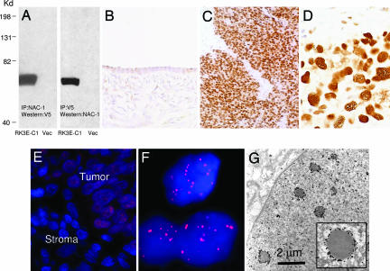
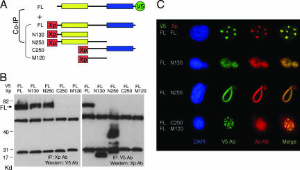
Recent studies have suggested an oncogenic role of the BTB/POZ-domain genes in hematopoietic malignancy. The aim of this study is to identify and characterize BTB/POZ-domain genes in the development of human epithelial cancers, i.e., carcinomas. In this study, we focused on ovarian carcinoma and analyzed gene expression levels using the serial analysis of gene expression (SAGE) data in all 130 deduced BTB/POZ genes. Our analysis reveals that NAC-1 is significantly overexpressed in ovarian serous carcinomas and several other types of carcinomas. Immunohistochemistry studies in ovarian serous carcinomas demonstrate that NAC-1 is localized in discrete nuclear bodies (tentatively named NAC-1 bodies), and the levels of NAC-1 expression correlate with tumor recurrence. Furthermore, intense NAC-1 immunoreactivity in primary tumors predicts early recurrence in ovarian cancer. Both coimmunoprecipitation and double immunofluorescence staining demonstrate that NAC-1 molecules homooligomerize through the BTB/POZ domain. Induced expression of the NAC-1 mutant containing only the BTB/POZ domain disrupts NAC-1 bodies, prevents tumor formation, and promotes tumor cell apoptosis in established tumors in a mouse xenograft model. Overexpression of full-length NAC-1 enhanced tumorigenicity of ovarian surface epithelial cells and NIH 3T3 cells in athymic nu/nu mice. In summary, NAC-1 is a tumor recurrence-associated gene with oncogenic potential, and the interaction between BTB/POZ domains of NAC-1 proteins is critical to form the discrete NAC-1 nuclear bodies and essential for tumor cell proliferation and survival.


Conflict of interest statement

The authors declare no conflict of interest.

Figures

Fig. 1.
Fig. 1.
Scatter plot of NAC-1 tags in several major tumor types. NAC-1 expression level is analyzed by counting NAC-1-specific tags from SAGE libraries in both cancer tissue (T, filled symbols) and the corresponding normal tissues (N, open symbols). The NAC-1 tags are normalized to tags per 100,000 (y axis). The dash line in each tumor type indicates the “ceiling” tag number in normal tissue libraries. Each symbol represents an individual specimen.
Fig. 2.
Fig. 2.
Immunoreactivity of NAC-1 in ovarian cancer tissues. (A) Immunoprecipitation/Western blot analyses using NAC-1 and V5 antibodies in RK3E cells transfected with pCDNA6-NAC-1/V5 (RK3E-C1) or vector-only control (Vec). A discrete band corresponding to NAC-1 protein mass is identified in this reciprocal analysis. (BD) The NAC-1 immunointensity is undetectable or weak in normal ovarian surface epithelium (B) but is strong in a high-grade serous carcinoma (C and D). (E and F) Immunofluorescence of NAC-1 protein localization in ovarian cancer cells in a tissue section. Tumor cells contain NAC-1 protein, which is located in discrete nuclear bodies (E). The adjacent stromal cells are negative for NAC-1 immunoreactivity. A higher magnification demonstrates the NAC-1 nuclear bodies by using a confocal fluorescence microscope (F). (G) Ultrastructure of NAC-1 bodies. ImmunoGold labeling of NAC-1-expressing RK3E cells demonstrates electron-dense bodies decorated by gold particles in the nuclear matrix.
Fig. 3.
Fig. 3.
NAC1 expression correlates with tumor progression in ovarian serous carcinomas. (A) Quantitative real-time PCR analysis shows higher NAC-1 expression levels in high-grade carcinomas (HG) than in ovarian surface epithelial cells (OSE), low-grade carcinomas (LG), and cystadenomas. Moreover, recurrent carcinomas have significantly higher expression levels than primary tumors (P = 0.012). The data are expressed as fold increase as compared with the average of OSE. (B) Immunohistochemistry demonstrates intense immunoreactivity in recurrent tumors as compared with patients' primary tumors in three representative cases.
Fig. 4.
Fig. 4.
Coimmunoprecipitation and colocalization of NAC-1 deletion mutants and full-length NAC-1. (A) Diagram of NAC-1 and NAC-1 deletion mutants. Full-length (FL) construct contains V5 tag at the C terminus, whereas all of the deletion mutants contain an Xpress (Xp) tag at the N terminus. The yellow box is the BTB/POZ domain; the blue box is the DUF1172 domain. (B) Coimmunoprecipitation shows that full-length NAC-1, N130, and N250 bind to NAC-1. The predicted molecular masses, not including the tag sequences, are: full-length NAC-1 (57.3 kDa), N130 (14.4 kDa), N250 (27.8 kDa), C250 (30.3 kDa), and M120 (14 kDa). (C) Cells with stable full-length NAC-1/V5 expression were transfected with different deletion mutants with the Xp tag. Double immunofluorescence shows that full-length NAC-1, N130, and N250 deletion mutants colocalize with full-length NAC-1. However, only full-length NAC-1 proteins form discrete round and oval-shaped NAC-1 nuclear bodies, whereas both N130 and N250 form irregular aggregates with the full-length NAC-1. Neither C250 nor M120 colocalizes with the full-length NAC-1 protein.
Fig. 5.
Fig. 5.
Effects of N130 induction on cellular proliferation and apoptosis in 96-well plates. (A and B) Cell growth curves show that after induction of N130 (−Dox), cell growth is significantly suppressed as compared with the noninduced cells (+Dox). In contrast, induction of C250 does not have an apparent effect on cell growth in SKOV3 (A) or HeLa (B) cells. (C) Cell cycle analysis shows an increase in G2/M fraction in N130-induced HeLa cells (Lower) as compared with noninduced cells (Upper) 24 h after induction, indicating a G2/M block. (D) Percentages of apoptotic and proliferating cells are determined by counting annexin V- and BrdU-positive cells, respectively, in both N130-induced and -noninduced cells. Data are presented as mean + SD. ∗, P < 0.05; ∗∗, P < 0.001, ∗∗∗, P < 0.0001, Student's t test.
Fig. 6.
Fig. 6.
Effects of constitutive expression of NAC-1 in immortalized ovarian surface epithelial cells (MOSE) and NIH 3T3 cells. Western blot analysis shows NAC-1 expression in stable clones of NAC-1-expressing MOSE cells (A) and NIH 3T3 cells (D). Upper blots, NAC-1; lower blots, GAPDH. Growth curves show a higher proliferation activity in both NAC-1 clones as compared with vector-transfected control under a low serum (0.5%) culture condition in MOSE cells (A) and NIH 3T3 cells (D). The weights of s.c. tumors increase in NAC-1-expressing MOSE tumors as compared with control MOSE in nude mice (B). A representative photomicrograph shows a s.c. NAC-1-expressing MOSE tumor (C). Similarly, the combined tumor weights of tumors in the peritoneal wall of the NAC-1-expressing NIH 3T3 cells are greater than the controls (E). (F) Cell proliferation was determined by a BrdU-incorporation assay, and all NAC-1-expressing clones have a higher proliferation rate than the vector-only control. Data are presented as mean + SD.

References

    1. Stogios PJ, Downs GS, Jauhal JJ, Nandra SK, Prive GG. Genome Biol. 2005;6:R82.1–R82.17. - PMC - PubMed
    1. Bardwell VJ, Treisman R. Genes Dev. 1994;8:1664–1677. - PubMed
    1. Albagli O, Dhordain P, Deweindt C, Lecocq G, Leprince D. Cell Growth Differ. 1995;6:1193–1198. - PubMed
    1. Chen Z, Brand NJ, Chen A, Chen SJ, Tong JH, Wang ZY, Waxman S, Zelent A. EMBO J. 1993;12:1161–1167. - PMC - PubMed
    1. Polo JM, Dell'Oso T, Ranuncolo SM, Cerchietti L, Beck D, Da Silva GF, Prive GG, Licht JD, Melnick A. Nat Med. 2004;10:1329–1335. - PubMed

Publication types

MeSH terms

Associated data