Esophageal perforation caused by fish vertebra ingestion in a seven-month-old infant demanded surgical intervention: A case report
- PMID: 17131491
- PMCID: PMC4087790
- DOI: 10.3748/wjg.v12.i44.7213
Esophageal perforation caused by fish vertebra ingestion in a seven-month-old infant demanded surgical intervention: A case report
Abstract
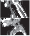
A seven-month-old infant was admitted to our hospital with a 1-wk history of shortness of breath, dysphagia, and fever. Diagnosis of esophageal perforation following fish vertebra ingestion was made by history review, pneumomediastinum and an irregular hyperdense lesion noted in initial chest radiogram. Neck computed tomography (CT) confirmed that the foreign body located at the cricopharyngeal level and a small esophageal tracheal fistula was shown by esophagogram. The initial response to treatment of fish bone removal guided by panendoscopy and antibiotics administration was poor since pneumothorax plus empyema developed. Fortunately, the patient's condition finally improved after decortication, mediastinotomy and perforated esophagus repair. To our knowledge, this is the first case report of esophageal perforation due to fish bone ingestion in infancy. In addition to particular caution that has to be taken when feeding the innocent, young victim, it may indicate the importance of surgical intervention for complicated esophageal perforation in infancy.
Figures


Similar articles
-
Successful endoscopic band ligation of esophageal perforation by fish bone ingestion.J Laparoendosc Adv Surg Tech A. 2013 May;23(5):459-62. doi: 10.1089/lap.2013.0082. Epub 2013 Apr 6. J Laparoendosc Adv Surg Tech A. 2013. PMID: 23560657
-
Esophageal perforation and mediastinitis from fish bone ingestion.South Med J. 2003 May;96(5):516-20. doi: 10.1097/01.SMJ.0000047744.34423.0B. South Med J. 2003. PMID: 12911196
-
Image of the month: Severe Mediastinitis and Pneumomediastinum Following Esophageal Perforation by a Fish Bone.Am J Gastroenterol. 2015 Sep;110(9):1262. doi: 10.1038/ajg.2015.68. Am J Gastroenterol. 2015. PMID: 26348300 No abstract available.
-
[Esophageal Foreign Body: Treatment and Complications].Korean J Gastroenterol. 2018 Jul 25;72(1):1-5. doi: 10.4166/kjg.2018.72.1.1. Korean J Gastroenterol. 2018. PMID: 30049171 Review. Korean.
-
Esophageal emergencies: WSES guidelines.World J Emerg Surg. 2019 May 31;14:26. doi: 10.1186/s13017-019-0245-2. eCollection 2019. World J Emerg Surg. 2019. PMID: 31164915 Free PMC article. Review.
Cited by
-
Uncommon Presentation of an Unusual Foreign Body.Indian J Crit Care Med. 2017 Jul;21(7):460-462. doi: 10.4103/ijccm.IJCCM_436_16. Indian J Crit Care Med. 2017. PMID: 28808368 Free PMC article.
-
A peculiar foreign body ingestion in 2-year-old girl complicated by esophageal perforation: case report and review of the literature.Oxf Med Case Reports. 2024 May 20;2024(5):omae040. doi: 10.1093/omcr/omae040. eCollection 2024 May. Oxf Med Case Reports. 2024. PMID: 38784778 Free PMC article.
-
Repeated colon penetration by an ingested fish bone: report of a case.Surg Today. 2008;38(4):363-5. doi: 10.1007/s00595-007-3629-y. Epub 2008 Mar 27. Surg Today. 2008. PMID: 18368330
-
Application of Three-dimensional Reconstruction in Esophageal Foreign Bodies.Korean J Thorac Cardiovasc Surg. 2011 Oct;44(5):368-72. doi: 10.5090/kjtcs.2011.44.5.368. Epub 2011 Oct 6. Korean J Thorac Cardiovasc Surg. 2011. PMID: 22263191 Free PMC article.
References
-
- Lai AT, Chow TL, Lee DT, Kwok SP. Risk factors predicting the development of complications after foreign body ingestion. Br J Surg. 2003;90:1531–1535. - PubMed
-
- Arana A, Hauser B, Hachimi-Idrissi S, Vandenplas Y. Management of ingested foreign bodies in childhood and review of the literature. Eur J Pediatr. 2001;160:468–472. - PubMed
-
- Uyemura MC. Foreign body ingestion in children. Am Fam Physician. 2005;72:287–291. - PubMed
-
- Jaffé A, Balfour-Lynn IM. Management of empyema in children. Pediatr Pulmonol. 2005;40:148–156. - PubMed
-
- Dahshan A. Management of ingested foreign bodies in children. J Okla State Med Assoc. 2001;94:183–186. - PubMed
Publication types
MeSH terms
LinkOut - more resources
Full Text Sources
Medical

