

Lentivirus-mediated silencing of Tiam1 gene influences multiple functions of a human colorectal cancer cell line
- PMID: 17132223
- PMCID: PMC1716017
- DOI: 10.1593/neo.06364
Lentivirus-mediated silencing of Tiam1 gene influences multiple functions of a human colorectal cancer cell line
Abstract
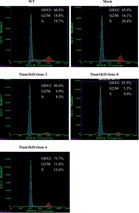
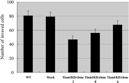
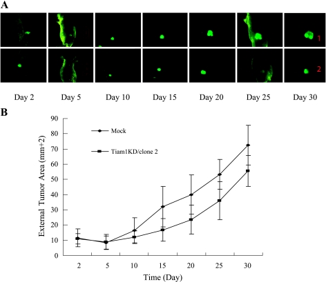
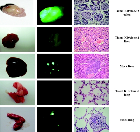
T lymphoma invasion and metastasis 1 (Tiam1) is a metastasis-related gene of T lymphoma that is also involved in the metastasis of a variety of other cancers. In this study, we tested the hypothesis that Tiam1 is a determinant of proliferation and metastasis in colorectal cancer, and we examined the effect of the inhibition of Tiam1 expression on proliferation and metastasis. We succeeded in establishing the Tiam1 knockdown colorectal cancer cell line using human immunodeficiency virus lentivirus-mediated RNA interference (RNAi) and found that the silencing of Tiam1 resulted in the effective inhibition of in vitro cell growth and of the invasive ability of colorectal cancer cells. Using an orthotopic xenograft model in nude mice, we confirmed that Tiam1 silencing could reduce tumor growth by subcutaneous injection and could suppress lung and liver metastases of colorectal cancer cells. Our results suggest that Tiam1 truly plays a causal role in the metastasis of colorectal cancer and that RNAi-mediated silencing of Tiam1 may provide an opportunity to develop a new treatment strategy for colorectal cancer.
Figures
Similar articles
-
Proteomic analysis of Tiam1-mediated metastasis in colorectal cancer.Cell Biol Int. 2007 Aug;31(8):805-14. doi: 10.1016/j.cellbi.2007.01.014. Epub 2007 Jan 21. Cell Biol Int. 2007. PMID: 17376711
-
The guanine nucleotide exchange factor Tiam1 increases colon carcinoma growth at metastatic sites in an orthotopic nude mouse model.Oncogene. 2005 Apr 7;24(15):2568-73. doi: 10.1038/sj.onc.1208503. Oncogene. 2005. PMID: 15735692
-
Inhibition of laryngeal cancer cell invasion and growth with lentiviral-vector delivered short hairpin RNA targeting human MMP-9 gene.Cancer Invest. 2008 Dec;26(10):984-9. doi: 10.1080/07357900802072897. Cancer Invest. 2008. PMID: 19093256
-
Suppression of growth of pancreatic cancer cell and expression of vascular endothelial growth factor by gene silencing with RNA interference.J Dig Dis. 2008 Nov;9(4):228-37. doi: 10.1111/j.1751-2980.2008.00352.x. J Dig Dis. 2008. PMID: 18959596
-
Lentivirus-mediated RNA interference targeting WWTR1 in human colorectal cancer cells inhibits cell proliferation in vitro and tumor growth in vivo.Oncol Rep. 2012 Jul;28(1):179-85. doi: 10.3892/or.2012.1751. Epub 2012 Apr 2. Oncol Rep. 2012. PMID: 22470139
Cited by
-
Increases in c-Yes expression level and activity promote motility but not proliferation of human colorectal carcinoma cells.Neoplasia. 2007 Sep;9(9):745-54. doi: 10.1593/neo.07442. Neoplasia. 2007. PMID: 17898870 Free PMC article.
-
miR-29b suppresses tumor growth and metastasis in colorectal cancer via downregulating Tiam1 expression and inhibiting epithelial-mesenchymal transition.Cell Death Dis. 2014 Jul 17;5(7):e1335. doi: 10.1038/cddis.2014.304. Cell Death Dis. 2014. PMID: 25032858 Free PMC article.
-
Tiam1 high expression is associated with poor prognosis in solid cancers: A meta-analysis.Medicine (Baltimore). 2019 Nov;98(45):e17529. doi: 10.1097/MD.0000000000017529. Medicine (Baltimore). 2019. PMID: 31702612 Free PMC article.
-
Adipogenic differentiation is not influenced by lentivirus-mediated shRNA targeting the SOCS3 gene in adipose-derived stromal cells.Mol Biol Rep. 2010 Jun;37(5):2455-62. doi: 10.1007/s11033-009-9757-2. Epub 2009 Aug 20. Mol Biol Rep. 2010. PMID: 19693688
-
Expression of Tiam1 predicts lymph node metastasis and poor survival of lung adenocarcinoma patients.Diagn Pathol. 2014 Mar 24;9:69. doi: 10.1186/1746-1596-9-69. Diagn Pathol. 2014. PMID: 24661909 Free PMC article.
References
-
- Haeusler LC, Blumenstein L, Stege P, Dvorsky R, Ahmadian MR. Comparative functional analysis of the Rac GTPases. FEBS Lett. 2003;555(3):556–560. - PubMed
-
- Michiels F, Collard JG. Rho-like GTPases: their role in cell adhesion and invasion. Biochem Soc Symp. 1999;65:125–146. - PubMed
-
- Denicola G, Tuveson DA. VAV1: a new target in pancreatic cancer? Cancer Biol Ther. 2005;4(5):509–511. - PubMed
-
- Perrot V, Vazquez-Prado J, Gutkind JS. Plexin B regulates Rho through the guanine nucleotide exchange factors leukemia-associated Rho GEF (LARG) and PDZ-RhoGEF. J Biol Chem. 2002;277(45):43115–43120. - PubMed
-
- Bassermann F, Jahn T, Miething C, Seipel P, Bai RY, Coutinho S, Tybulewicz VL, Peschel C, Duyster J. Association of Bcr-Abl with the proto-oncogene Vav is implicated in activation of the Rac-1 pathway. J Biol Chem. 2002;277(14):12437–12445. - PubMed
Publication types
MeSH terms
Substances
LinkOut - more resources
Full Text Sources
Medical