

Effect of hyperosmolality on beta-defensin gene expression by human corneal epithelial cells
- PMID: 17133055
- PMCID: PMC2430508
- DOI: 10.1097/01.ico.0000228785.84581.35
Effect of hyperosmolality on beta-defensin gene expression by human corneal epithelial cells
Abstract
Purpose: As human beta-defensins (hBD) are important antimicrobial peptides at epithelial surfaces, including the ocular surface, we tested the effect of hyperosmolar conditions on the expression of these peptides by human corneal epithelial cells (HCECs).
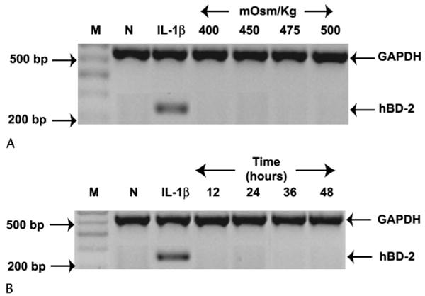
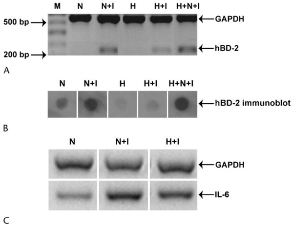
Methods: Simian virus 40-transformed HCECs (n = 5) or primary cultured HCECs (n = 5) were treated with serum-free media or serum-free hyperosmolar (400-500 mOsm/kg) media for 24 hours or serum-free 500 mOsm/kg media for 12 to 48 hours. The effect of hyperosmolality on interleukin-1beta (IL-1beta)-induced hBD-2 expression was also tested. IL-6 expression was studied as a marker of IL-1beta function. Expression of hBD-1, -2, and -3 and IL-6 mRNA was detected by reverse transcription-polymerase chain reaction (RT-PCR). The levels of active IL-1beta (culture supernatants and cell lysates) and pro-IL-1beta (cell lysates) were detected by enzyme-linked immunosorbent assay.
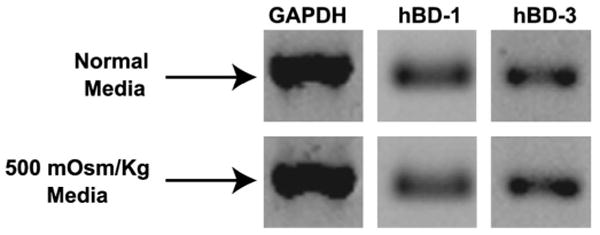
Results: HCECs constitutively expressed hBD-1 and -3 but not hBD-2. Hyperosmolar media had no effect on the basal expression of hBD-1 or -3 and did not induce the expression of hBD-2. Treatment with 500 mOsm/kg media for 24 hours decreased the ability of IL-1beta to upregulate hBD-2 and IL-6 expression. Active or pro-IL-1beta was not detected in any cell culture sample.
Conclusion: Our results suggest that the hyperosmolar environment observed in diseases such as dry eye does not alter defensin expression. However, a hyperosmolar environment may influence cytokine function in ocular surface cells and thus affect their response to injury and inflammation.
Figures
Similar articles
-
Defensin expression by the cornea: multiple signalling pathways mediate IL-1beta stimulation of hBD-2 expression by human corneal epithelial cells.Invest Ophthalmol Vis Sci. 2003 May;44(5):1859-65. doi: 10.1167/iovs.02-0787. Invest Ophthalmol Vis Sci. 2003. PMID: 12714616 Free PMC article.
-
Toll-like receptor activation modulates antimicrobial peptide expression by ocular surface cells.Exp Eye Res. 2011 Mar;92(3):209-20. doi: 10.1016/j.exer.2010.12.005. Epub 2010 Dec 31. Exp Eye Res. 2011. PMID: 21195713 Free PMC article.
-
Ocular surface expression and in vitro activity of antimicrobial peptides.Curr Eye Res. 2007 Jul-Aug;32(7-8):595-609. doi: 10.1080/02713680701446653. Curr Eye Res. 2007. PMID: 17852183 Free PMC article.
-
Expression of human beta-defensins in conjunctival epithelium: relevance to dry eye disease.Invest Ophthalmol Vis Sci. 2003 Sep;44(9):3795-801. doi: 10.1167/iovs.02-1301. Invest Ophthalmol Vis Sci. 2003. PMID: 12939294 Free PMC article.
-
Effects of L-carnitine, erythritol and betaine on pro-inflammatory markers in primary human corneal epithelial cells exposed to hyperosmotic stress.Curr Eye Res. 2015 Jul;40(7):657-67. doi: 10.3109/02713683.2014.957776. Epub 2014 Oct 1. Curr Eye Res. 2015. PMID: 25271595 Free PMC article.
Cited by
-
Mucosal immunology of the ocular surface.Mucosal Immunol. 2022 Jun;15(6):1143-1157. doi: 10.1038/s41385-022-00551-6. Epub 2022 Aug 24. Mucosal Immunol. 2022. PMID: 36002743 Free PMC article. Review.
-
Role of Pattern Recognition Receptors in the Modulation of Antimicrobial Peptide Expression in the Corneal Epithelial Innate Response to F. solani.Invest Ophthalmol Vis Sci. 2017 May 1;58(5):2463-2472. doi: 10.1167/iovs.16-20658. Invest Ophthalmol Vis Sci. 2017. PMID: 28460048 Free PMC article.
-
Tear cytokine profile as a noninvasive biomarker of inflammation for ocular surface diseases: standard operating procedures.Invest Ophthalmol Vis Sci. 2013 Dec 23;54(13):8327-36. doi: 10.1167/iovs.13-12132. Invest Ophthalmol Vis Sci. 2013. PMID: 24204044 Free PMC article. Clinical Trial.
-
In Vitro Inhibition of NFAT5-Mediated Induction of CCL2 in Hyperosmotic Conditions by Cyclosporine and Dexamethasone on Human HeLa-Modified Conjunctiva-Derived Cells.PLoS One. 2016 Aug 3;11(8):e0159983. doi: 10.1371/journal.pone.0159983. eCollection 2016. PLoS One. 2016. PMID: 27486749 Free PMC article.
-
Effect of different artificial tears against desiccation in cultured human epithelial cells.Med Sci Monit. 2012 May;18(5):BR188-92. doi: 10.12659/msm.882728. Med Sci Monit. 2012. PMID: 22534701 Free PMC article.
References
-
- Gilbard JP, Farris RL, Santamaria J., 2nd Osmolarity of tear microvolumes in keratoconjunctivitis sicca. Arch Ophthalmol. 1978;96:677–681. - PubMed
-
- Farris RL. Tear osmolarity—a new gold standard? Adv Exp Med Biol. 1994;350:495–503. - PubMed
-
- Bron AJ, Tiffany JM, Yokoi N, et al. Using osmolarity to diagnose dry eye: a compartmental hypothesis and review of our assumptions. Adv Exp Med Biol. 2002;506:1087–1095. - PubMed
-
- Anagnoste SR, Hall LS, Lisman RD. Vapor pressure osmolarity of tears as a clinical tool for evaluation of keratoconjunctivitis sicca. Invest Ophthalmol Vis Sci. 1996;37:S852.
-
- Gilbard JP, Rossi SR, Heyda KG. Ophthalmic solutions, the ocular surface, and a unique therapeutic artificial tear formulation. Am J Ophthalmol. 1989;107:348–355. - PubMed
Publication types
MeSH terms
Substances
Grants and funding
LinkOut - more resources
Full Text Sources
Research Materials
