Hemodynamic responses in neural circuitries for detection of visual target and novelty: An event-related fMRI study
- PMID: 17133387
- PMCID: PMC6871418
- DOI: 10.1002/hbm.20319
Hemodynamic responses in neural circuitries for detection of visual target and novelty: An event-related fMRI study
Abstract
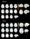
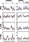
The oddball paradigm examines attentional processes by establishing neural substrates for target detection and novelty. Event-related functional imaging enables characterization of hemodynamic changes associated with these processes. We studied 36 healthy participants (17 men) applying a visual oddball event-related design at 4 Tesla, and performed an unbiased determination of the hemodynamic response function (HRF). Targets were associated with bilateral, albeit leftward predominant changes in frontal-parietal temporal and occipital cortices, and limbic and basal ganglia regions. Activation to novelty was more posteriorly distributed, and frontal activation occurred only on the right, while robust activation was seen in occipital regions bilaterally. Overlapping regions were left thalamus, caudate and cuneus and right parietal precuneus. While robust HRFs characterized most regions, target detection was associated with a negative HRF in the right parietal precuneus and a biphasic HRF in thalamus, basal ganglia, and all occipital regions. Both height of the HRF and longer time to peak in the right cingulate were associated with slower response time. Sex differences were observed, with higher HRF peaks for novelty in men in right occipital regions, and longer time to peak in the left hemisphere. Age was associated with reduced peak HRF in left frontal region. Thus, indices of the HRF can be used to better understand the relationship between hemodynamic changes and performance and can be sensitive to individual differences.
(c) 2006 Wiley-Liss, Inc.
Figures


Similar articles
-
Variation of BOLD hemodynamic responses across subjects and brain regions and their effects on statistical analyses.Neuroimage. 2004 Apr;21(4):1639-51. doi: 10.1016/j.neuroimage.2003.11.029. Neuroimage. 2004. PMID: 15050587
-
Visual attention circuitry in schizophrenia investigated with oddball event-related functional magnetic resonance imaging.Am J Psychiatry. 2007 Mar;164(3):442-9. doi: 10.1176/ajp.2007.164.3.442. Am J Psychiatry. 2007. PMID: 17329469
-
Responses to rare visual target and distractor stimuli using event-related fMRI.J Neurophysiol. 2000 May;83(5):3133-9. doi: 10.1152/jn.2000.83.5.3133. J Neurophysiol. 2000. PMID: 10805707 Clinical Trial.
-
Cerebral correlates of alerting, orienting and reorienting of visuospatial attention: an event-related fMRI study.Neuroimage. 2004 Jan;21(1):318-28. doi: 10.1016/j.neuroimage.2003.08.044. Neuroimage. 2004. PMID: 14741670 Clinical Trial.
-
Control networks and hemispheric asymmetries in parietal cortex during attentional orienting in different spatial reference frames.Neuroimage. 2005 Apr 15;25(3):668-83. doi: 10.1016/j.neuroimage.2004.07.075. Neuroimage. 2005. PMID: 15808968
Cited by
-
Neural correlates of the perception for novel objects.PLoS One. 2013 Apr 30;8(4):e62979. doi: 10.1371/journal.pone.0062979. Print 2013. PLoS One. 2013. PMID: 23646167 Free PMC article.
-
The White Matter Microintegrity Alterations of Neocortical and Limbic Association Fibers in Major Depressive Disorder and Panic Disorder: The Comparison.Medicine (Baltimore). 2016 Mar;95(9):e2982. doi: 10.1097/MD.0000000000002982. Medicine (Baltimore). 2016. PMID: 26945417 Free PMC article.
-
Decreasing predictability of visual motion enhances feed-forward processing in visual cortex when stimuli are behaviorally relevant.Brain Struct Funct. 2017 Mar;222(2):849-866. doi: 10.1007/s00429-016-1251-8. Epub 2016 Jun 22. Brain Struct Funct. 2017. PMID: 27334340 Free PMC article.
-
Neuropharmacological effect of methylphenidate on attention network in children with attention deficit hyperactivity disorder during oddball paradigms as assessed using functional near-infrared spectroscopy.Neurophotonics. 2014 Jul;1(1):015001. doi: 10.1117/1.NPh.1.1.015001. Epub 2014 May 28. Neurophotonics. 2014. PMID: 26157971 Free PMC article.
-
Direction and magnitude of nicotine effects on the fMRI BOLD response are related to nicotine effects on behavioral performance.Psychopharmacology (Berl). 2011 May;215(2):333-44. doi: 10.1007/s00213-010-2145-8. Epub 2011 Jan 18. Psychopharmacology (Berl). 2011. PMID: 21243486 Free PMC article. Clinical Trial.
References
-
- Andersson JL ( 1997): How to estimate global activity independent of changes in local activity. Neuroimage 6: 237–244. - PubMed
-
- Ardekani BA, Choi SJ, Hossein‐Zadeh GA, Porjesz B, Tanabe JL, Lim KO, Bilder R, Helpern JA, Begleiter H 2002): Functional magnetic resonance imaging of brain activity in the visual oddball task. Brain Res Cogn Brain Res 14: 347–356. - PubMed
-
- Bahramali H, Gordon E, Lagopoulos J, Lim CL, Li W, Leslie J, Wright JJ ( 1999): The effects of age on late components of the ERP and reaction time. Exp Aging Res 26: 69–80. - PubMed
-
- Bledowski C, Prvulovic D, Goebel R, Zanella FE, Linden DE ( 2004): Attentional systems in target and distractor processing: A combined ERP and fMRI study. Neuroimage 22: 530–540. - PubMed
Publication types
MeSH terms
Grants and funding
LinkOut - more resources
Full Text Sources
Medical

