

Regulation of emotional responses elicited by threat-related stimuli
- PMID: 17133391
- PMCID: PMC6871321
- DOI: 10.1002/hbm.20291
Regulation of emotional responses elicited by threat-related stimuli
Abstract
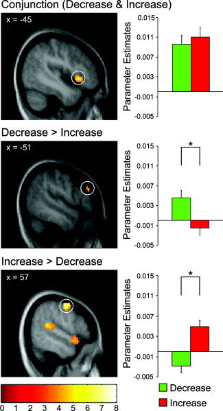
The capacity to voluntarily regulate emotions is critical for mental health, especially when coping with aversive events. Several neuroimaging studies of emotion regulation found the amygdala to be a target for downregulation and prefrontal regions to be associated with downregulation. To characterize the role of prefrontal regions in bidirectional emotion regulation and to investigate regulatory influences on amygdala activity and peripheral physiological measures, a functional magnetic resonance imaging (fMRI) study with simultaneous recording of self-report, startle eyeblink, and skin conductance responses was carried out. Subjects viewed threat-related pictures and were asked to up- and downregulate their emotional responses using reappraisal strategies. While startle eyeblink responses (in successful regulators) and skin conductance responses were amplified during upregulation, but showed no consistent effect during downregulation, amygdala activity was increased and decreased according to the regulation instructions. Trial-by-trial ratings of regulation success correlated positively with activity in amygdala during upregulation and orbitofrontal cortex during downregulation. Downregulation was characterized by left-hemispheric activation peaks in anterior cingulate cortex, dorsolateral prefrontal cortex, and orbitofrontal cortex and upregulation was characterized by a pattern of prefrontal activation not restricted to the left hemisphere. Further analyses showed significant overlap of prefrontal activation across both regulation conditions, possibly reflecting cognitive processes underlying both up- and downregulation, but also showed distinct activations in each condition. The present study demonstrates that amygdala responses to threat-related stimuli can be controlled through the use of cognitive strategies depending on recruitment of prefrontal areas, thereby changing the subject's affective state.
Figures


References
-
- Adcock RA, Lutomski K, McLeod SR, Soneji DJ, Gabriele JDE ( 2005): Real‐time fMRI during the psychotherapy session: toward a methodology to augment therapeutic benefit, exemplary data. Abstract Presented at the Human Brain Mapping Conference 2005, Toronto, Canada.
-
- Anand A, Li Y, Wang Y, Wu J, Gao S, Bukhari L, Mathews VP, Kalnin A, Lowe MJ ( 2005): Activity and connectivity of brain mood regulating circuit in depression: a functional magnetic resonance study. Biol Psychiatry 57: 1079–1088. - PubMed
-
- Anders S, Weiskopf N, Lule D, Birbaumer N ( 2004a): Infrared oculography—validation of a new method to monitor startle eyeblink amplitudes during fMRI. Neuroimage 22: 767–770. - PubMed
-
- Anderson AK, Christoff K, Stappen I, Panitz D, Ghahremani DG, Glover G, Gabrieli JD, Sobel N ( 2003): Dissociated neural representations of intensity and valence in human olfaction. Nat Neurosci 6: 196–202. - PubMed
Publication types
MeSH terms
Substances
LinkOut - more resources
Full Text Sources
Other Literature Sources
