

Second site escape of a T20-dependent HIV-1 variant by a single amino acid change in the CD4 binding region of the envelope glycoprotein
- PMID: 17134507
- PMCID: PMC1698932
- DOI: 10.1186/1742-4690-3-84
Second site escape of a T20-dependent HIV-1 variant by a single amino acid change in the CD4 binding region of the envelope glycoprotein
Abstract
Background: We previously described the selection of a T20-dependent human immunodeficiency virus type-1 (HIV-1) variant in a patient on T20 therapy. The fusion inhibitor T20 targets the viral envelope (Env) protein by blocking a conformational switch that is critical for viral entry into the host cell. T20-dependent viral entry is the result of 2 mutations in Env (GIA-SKY), creating a protein that undergoes a premature conformational switch, and the presence of T20 prevents this premature switch and rescues viral entry. In the present study, we performed 6 independent evolution experiments with the T20-dependent HIV-1 variant in the absence of T20, with the aim to identify second site compensatory changes, which may provide new mechanistic insights into Env function and the T20-dependence mechanism.
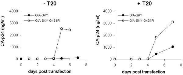
Results: Escape variants with improved replication capacity appeared within 42 days in 5 evolution cultures. Strikingly, 3 cultures revealed the same single amino acid change in the CD4 binding region of Env (glycine at position 431 substituted for arginine: G431R). This mutation was sufficient to abolish the T20-dependence phenotype and restore viral replication in the absence of T20. The GIA-SKY-G431R escape variant produces an Env protein that exhibits reduced syncytia formation and reduced cell-cell fusion activity. The escape variant was more sensitive to an antibody acting on an early gp41 intermediate, suggesting that the G431R mutation helps preserve a pre-fusion Env conformation, similar to T20 action. The escape variant was also less sensitive to soluble CD4, suggesting a reduced CD4 receptor affinity.
Conclusion: The forced evolution experiments indicate that the premature conformational switch of the T20-dependent HIV-1 Env variant (GIA-SKY) can be corrected by a second site mutation in Env (GIA-SKY-G431R) that affects the interaction with the CD4 receptor.
Figures


Similar articles
-
An anti-human immunodeficiency virus multiple antigen peptide encompassing the cleavage region of the env precursor interferes with membrane fusion at a post-CD4 binding step.Virology. 2000 Jul 20;273(1):169-77. doi: 10.1006/viro.2000.0368. Virology. 2000. PMID: 10891419
-
Emergence of a drug-dependent human immunodeficiency virus type 1 variant during therapy with the T20 fusion inhibitor.J Virol. 2004 Nov;78(22):12428-37. doi: 10.1128/JVI.78.22.12428-12437.2004. J Virol. 2004. PMID: 15507629 Free PMC article.
-
HIV-1 gp41 Residues Modulate CD4-Induced Conformational Changes in the Envelope Glycoprotein and Evolution of a Relaxed Conformation of gp120.J Virol. 2018 Jul 31;92(16):e00583-18. doi: 10.1128/JVI.00583-18. Print 2018 Aug 15. J Virol. 2018. PMID: 29875245 Free PMC article.
-
CD4 activation of HIV fusion.Int J Cell Cloning. 1992 Nov;10(6):323-32. doi: 10.1002/stem.5530100603. Int J Cell Cloning. 1992. PMID: 1281202 Review.
-
Potential drug targets on the HIV-1 envelope glycoproteins, gp120 and gp41.Curr Pharm Des. 2003;9(18):1453-62. doi: 10.2174/1381612033454720. Curr Pharm Des. 2003. PMID: 12769725 Review.
Cited by
-
Distinct efficacy of HIV-1 entry inhibitors to prevent cell-to-cell transfer of R5 and X4 viruses across a human placental trophoblast barrier in a reconstitution model in vitro.Retrovirology. 2008 Mar 31;5:31. doi: 10.1186/1742-4690-5-31. Retrovirology. 2008. PMID: 18377645 Free PMC article.
-
Dual-reporter phenotypic assay for human immunodeficiency viruses.J Clin Microbiol. 2008 Feb;46(2):792-5. doi: 10.1128/JCM.01470-07. Epub 2007 Dec 19. J Clin Microbiol. 2008. PMID: 18094135 Free PMC article.
-
HIV-1 evolution: frustrating therapies, but disclosing molecular mechanisms.Philos Trans R Soc Lond B Biol Sci. 2010 Jun 27;365(1548):1965-73. doi: 10.1098/rstb.2010.0072. Philos Trans R Soc Lond B Biol Sci. 2010. PMID: 20478891 Free PMC article. Review.
-
Design of peptide-based inhibitors for human immunodeficiency virus type 1 strains resistant to T-20.J Biol Chem. 2009 Feb 20;284(8):4914-20. doi: 10.1074/jbc.M807169200. Epub 2008 Dec 10. J Biol Chem. 2009. PMID: 19073606 Free PMC article.
-
The 2008 Retrovirology Prize: Ben Berkhout and his RNA world.Retrovirology. 2008 Dec 11;5:113. doi: 10.1186/1742-4690-5-113. Retrovirology. 2008. PMID: 19077224 Free PMC article.
References
Publication types
MeSH terms
Substances
LinkOut - more resources
Full Text Sources
Research Materials
