Comparative Study
. 2007 May;129(1-2):76-92.
doi: 10.1016/j.pain.2006.09.039. Epub 2006 Nov 28.

Immunohistochemical localization of histamine H3 receptors in rodent skin, dorsal root ganglia, superior cervical ganglia, and spinal cord: potential antinociceptive targets

Affiliations
Comparative Study

Immunohistochemical localization of histamine H3 receptors in rodent skin, dorsal root ganglia, superior cervical ganglia, and spinal cord: potential antinociceptive targets

Keri E Cannon et al. Pain. 2007 May.

Abstract

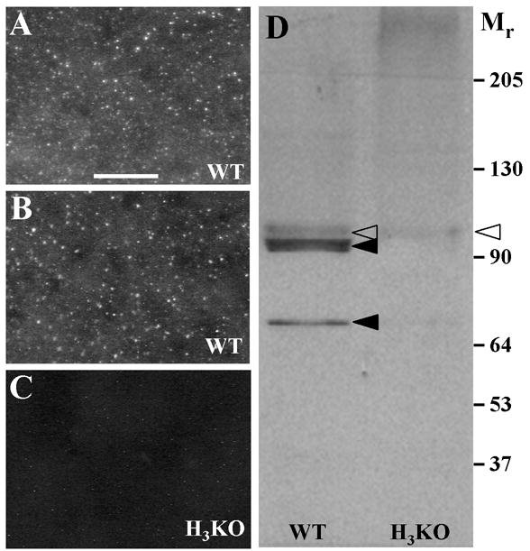
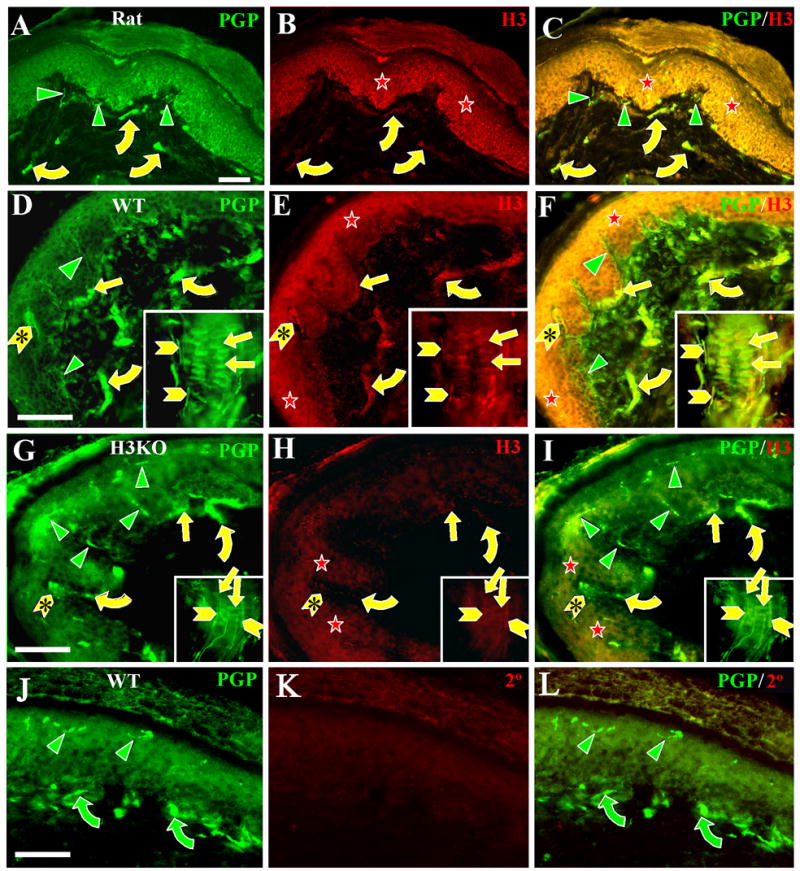
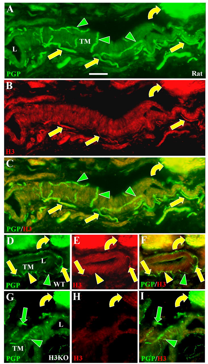
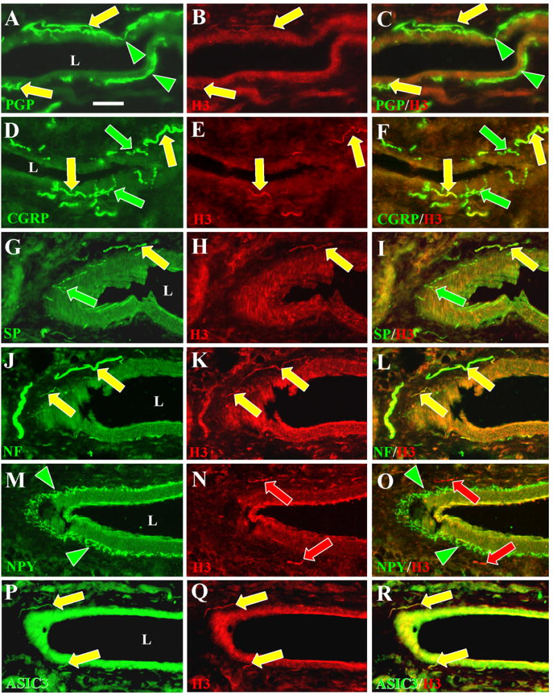
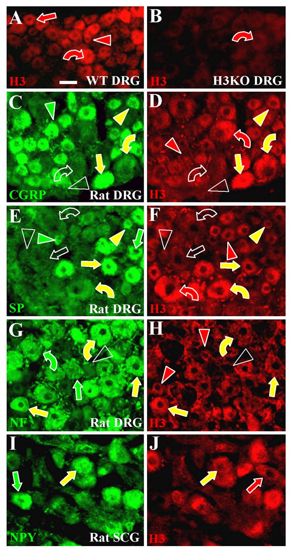
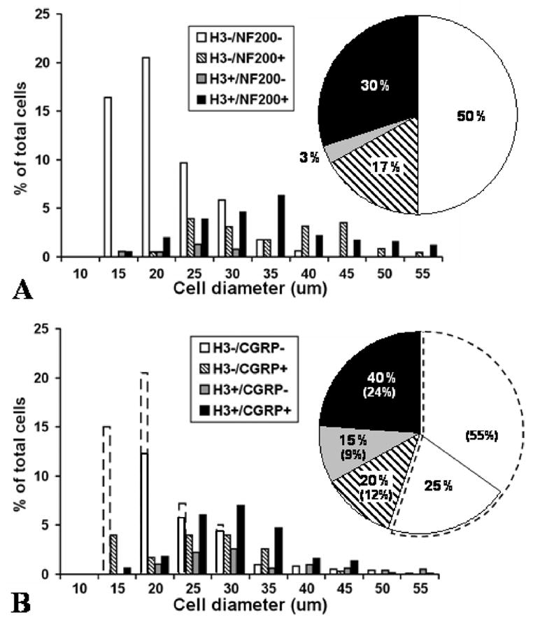
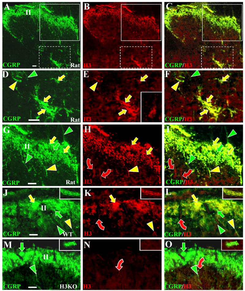
Activation of histamine H3 receptors (H3Rs) reduces inflammation and nociception, but the existence of H3Rs on peripheral innervation has never been demonstrated. Here we use antibodies to locate H3Rs in whisker pads, hairy and glabrous hind paw skin, dorsal root ganglia (DRGs), and spinal cords of rats, wild type mice, and H3R knockout (H3KO) mice. Although H3Rs have been hypothesized to be on C and sympathetic fibers, H3R-like immunoreactivity (H3R-LI) was only detected on presumptive periarterial A delta fibers and on A beta fibers that terminated in Meissner's corpuscles and as lanceolate endings around hair follicles. The H3R-positive periarterial fibers were thin-caliber and coexpressed immunoreactivity for calcitonin gene-related peptide (CGRP), substance P, acid sensing ion channel 3, and 200 kDa neurofilament protein (NF). H3R-LI was also detected on epidermal keratinocytes and Merkel cells, but not on Merkel endings, C fibers, any other A delta fibers, or sympathetic fibers. In DRGs, H3R-LI was preponderantly on medium to large neurons coexpressing NF-LI and mostly CGRP-LI. In dorsal horn, CGRP-positive fibers with and without H3R-LI ramified extensively in lamina II; many of the former formed a plexus in lamina V. Low levels of H3R-LI were also present on A beta fibers penetrating superficial and into deeper laminae. The distribution of H3R-LI was similar in rats and wild type mice, but was eliminated or strongly reduced in A delta fibers and A beta fibers, respectively, in H3KO mice. Taken with recently published behavioral results, the present findings suggest that periarterial, peptidergic, H3R-containing A delta fibers may be sources of high threshold mechanical nociception.


Figures

Figure 1
Figure 1. Characterization of anti H3R labeling
A–C. Digital images of punctate anti-H3R labeling among the neurons in the striatum as seen in coronal sections from wildtype (A, B) and H3KO mice (C). Scale bar, 25 μm. Comparable labeling was obtained with anti-H3R antibodies produced independently by Chazot et al. (2001) (A) and Chemicon (B). Virtually all labeling is absent in the striatum of H3KO mice prepared with the Chazot antibody (C). D. A Western blot with the Chazot antibody reveals two dense bands (solid arrowheads) in the wildtype mice that are eliminated in H3KO mice, and a faint band (open arrowheads) that is reduced but still persists in the H3KO mice.
Figure 2
Figure 2. H3R -LI is absent on thin caliber innervation to the epidermis and upper dermis, but is present on some Aβ fiber innervation, Merkel cells and keratinocytes
Digital images of glabrous hind paw sections from rats (A–C), wild type mice (D–F, J–L), and H3KO (G–I) mice double labeled with anti-PGP (left panels) and anti-H3R (middle panels). Merged double labeled images are shown in the right panels. Insets in D–I show labeling in whisker follicles from mystacial pads. Scale bars = 25 μm (50μm for insets). In this and subsequent figures, green (Cy2 or Alexa 488) and red (Cy3) symbols indicate single labeled structures; yellow symbols indicate double labeled structures. A–F: As seen in rat and wildtype mouse skin, thin caliber innervation labeled with anti-PGP (green arrowheads) in the epidermis or upper dermis is not labeled with anti-H3R. This includes all C-fiber and Aδ-fiber innervation. Anti-PGP and anti-H3R double labeling was present on some large caliber Aβ fibers (yellow curved arrows), and on Aβ-fiber endings in Meissner corpuscles (yellow chevrons with asterisks) and Aβ-fiber endings on whisker follicles (yellow chevrons) and hair follicles (not shown). H3R-LI is also present on keratinocytes in the epidermis (red stars) as well as on anti-PGP labeled Merkel cells (yellow straight arrows) in lamina basalis of the epidermis and the outer root sheath of whisker follicles. H3R-LI was not evident on the Aβ fibers that innervate the Merkel cells. G–I: In H3KO mice, H3R-LI is drastically reduced but faint residual Cy3 labeling was detected on keratinocytes in the epidermis (red stars), some Aβ fibers (yellow curved arrows), Meissner corpuscles (yellow chevrons with arrowheads), lanceolate endings (yellow chevrons) and Merkel cells (yellow straight arrows). All thin caliber innervation was Cy3 negative (green arrowheads). J–L. Images of wildtype glabrous mouse skin double labeled with anti-PGP and peptide preabsorbed anti-H3R. None of the innervation (green arrowheads and curved arrows) nor cells in the epidermis were labeled with the preabsorbed anti-H3R.
Figure 3
Figure 3. H3R-LI on periarterial innervation in the deep dermis
Digital images of glabrous hind paw sections from rats (A–C), wildtype mice (D–F), and H3KO mice (G–I) double labeled with anti-PGP (A, D, G) and anti H3R revealed with Cy3 (B, E, H). Scale bars, 25 μm. L = arterial lumens, TM = tunica media. A–F. Double labeling is present among individual axons and bundles of axons (yellow straight arrows) which are located in the tunica adventitia (which surrounds the tunica media) and have previously been shown to be mostly CGRP-positive presumptive C fibers and Aδ fibers (see also Fig. 4). Thin-caliber fibers that ramify at the interface between the tunica adventitia and tunica media have previously been shown to be NPY and TH-positive and are presumptive sympathetic innervation. They were entirely labeled only with anti-PGP (green arrowheads) in the rat, but some double labeled with anti-H3R in the mouse (yellow arrowheads). Many Aβ fibers in large deep dermal nerves were double labeled (yellow curved arrows). G–I. No H3R-LI was present among sensory (green arrows) and sympathetic (green arrowheads) periarterial fibers in H3KO mice, but faint residual labeling was detected on Aβ fibers (yellow curved arrows).
Figure 4
Figure 4. Co-localization of H3 receptors with CGRP, SP, NF, and ASIC3, but not NPY on deep dermal, peptidergic, arterial innervation
Digital images of glabrous hind paw sections from rats double labeled with antibodies against H3R (middle panels) and against several other neuronal antigens (left panels). Merged images are in the right panels. The Chemicon anti-H3R used in B (yellow arrows) did not label innervation as intensely as the Chazot antibody used in all the other middle panels (red and yellow arrows). Scale bar, 25 μm. L = lumen of arteries. A–C. H3R-LI was expressed almost entirely among the anti-PGP individual fibers and bundles of fibers (yellow arrows) located in the tunica adventitia which surrounds the tunica media. Anti-PGP labeled fibers that ramify at the interface between the tunica adventitia and tunica media lack H3R-LI (green arrowheads). D–F. CGRP-LI was expressed in the periarterial innervation that is presumably sensory (yellow and green arrows). A subset of this innervation co-expresses H3R–LI (yellow arrows). G–I. SP-LI was also expressed in periarterial fibers that are presumably sensory (yellow and green arrows). H3R-LI was expressed on a subset of this innervation (yellow arrows). J–L. H3R–LI was expressed on periarterial fibers that co-labeled with anti-NF. Previous studies showed that the NF-positive fibers are a subset of those that label with anti-CGRP. M–O. H3R-LI was only expressed on fibers (red arrows) that were distinct from those that label with anti-NPY (green arrowheads). P–R. H3R-positive fibers also labeled with anti-ASIC3 (yellow arrows).
Figure 5
Figure 5. Immunochemical characterization of H3R expressing neurons in lumbar dorsal root ganglia (DRG) and superior cervical ganglia (SCG)
Sections of ganglia were all labeled with anti-H3R (red) and some sections were double labeled with another antibodies (green). Small size neurons are indicated by arrowheads, medium size neurons by straight arrows and large size neurons by curved arrows. Red indicators show neurons only labeled with anti-H3R, green indicators only neurons labeled with other antibodies, and yellow indicators neurons that are double labeled. Empty indicators show neurons that were regarded as having only background fluorescence. Scale bar = 25μm. A, B. Images from mouse DRG sections. Anti-H3R labeled many of the small (red arrowhead), medium (red arrow) and large (red curved arrow) DRG neurons from a wildtype mouse (A). Anti-H3R is virtually eliminated in DRG neurons from a H3KO mouse (B) except for very low residual leveling on some relatively large neurons (red curved arrow). C–H. Images from rat lumbar DRG sections. C, D. Small neurons double labeled with anti-CGRP and anti-H3R (yellow arrowheads), only labeled with anti-CGRP (green arrowhead), or only labeled with anti-H3R (red arrowhead). Some small neurons were not labeled with either antibody (unfilled arrowheads). Most medium size neurons expressed both CGRP-IR and H3R-IR (yellow straight arrows). Large neurons were typically immunolabeled for both CGRP and H3R (yellow curved arrows) or were negative for both (unfilled curved arrows). E, F. Overall, fewer neurons label with anti-SP than with anti-CGRP. Some small neurons were double labeled with anti-SP and anti-H3R (yellow arrowheads), only labeled with anti-SP (green arrowhead), or only labeled with anti-H3R (red arrowhead). Some small neurons were not labeled with either antibody (unfilled arrowheads). Most medium size neurons were immunolabeled for both SP and H3R (yellow arrows) or negative for both (unfilled arrow). The green straight arrow indicates a relatively rare medium size neuron that only expressed SP-IR. Some large neurons were double labeled with anti-SP and anti-H3R (yellow curved arrows), some were labeled only with anti-H3R (red curved arrow), and others were not labeled with either antibody (unfilled curved arrows). G, H. Virtually all small neurons were NF-negative, whereas virtually all medium and large neurons were NF-positive. Small neurons were H3R-positive (red arrowheads) or negative (unfilled arrowheads). Medium and large neurons were H3R-positive (yellow straight arrows and curved arrows) or H3R -negative (green straight arrows and curved arrows). I, J. Double labeling of rat superior cervical ganglia revealed neurons that coexpressed anti-NPY-IR and anti-H3-IR (yellow arrows) or that are positive only for NPY (green arrow) or H3R (red arrow).
Figure 6
Figure 6. Quantification of anti-H3R labeling in rat dorsal root ganglia
Distribution of cell sizes and labeling characteristics in rat lumbar DRG neurons as assessed in sections double labeled with anti-H3R and anti-NF (A) or with anti-H3R and anti-CGRP (B). Data were normalized from measurements of 758 cells in three L4 and L5 rat DRGs and the proportions were corrected for cell size. A. After double labeling with anti-H3R and anti-NF, 33% of all presumptive neurons had H3R–LI. Most of these H3R–LI neurons were NF-positive (30% vs. 3%) and had medium to large diameters. 17% of all detected neurons were NF-positive and H3R-negative and had a bimodal distribution among medium and large cells. 50% of presumptive neurons were negative for both H3R-LI and NF-LI, and most had relatively small diameters. B. After double labeling with anti-H3R and anti-CGRP, the actual detected proportions of H3R-positive and negative neurons are shown in the solid line enclosed bars and pie chart sectors. The actual percentages of double-labeled, single-labeled and unlabeled neurons are shown by the numbers without brackets in the pie chart sectors. The proportion of anti-H3R labeled neurons in B was much higher (55%) than in A (33%), but presumably should be the same. This was most likely due to a failure to detect many unlabeled cells in the anti-H3R and anti-CGRP labeled sections (see Methods and Results). To correct for this likely lack of detection, the total % of H3R–LI neurons observed in the anti-H3R and anti-CGRP (B) was normalized to 33% by adjusting the proportion of H3R-negative/CGRP-negative cells to reflect the level of H3R-negative neurons observed in the anti-H3R and anti-NF labeled sections (A). The adjusted increase is indicated by broken line bars and pie sectors, and the adjusted percentages are the numbers shown in brackets in B. With or without this adjustment, neurons with H3R–LI had predominantly medium to large diameters, with a 3:1 ratio of CGRP-positive to CGRP-negative neurons (24% vs 9% in adjusted percentages). Based on adjusted percentages, 12% of the neurons were H3R-negative and CGRP-positive and had a bimodal distribution among small and medium diameters. Even without an adjustment, the neurons that lacked both H3R–LI and CGRP-LI were skewed towards smaller diameters (white bars and white pie sector within solid lines).
Figure 7
Figure 7. Anti-H3R labeling in the dorsal horn of rats, wild type mice, and H3KO mice
Digital cross sectional images of lumbar spinal cords from rats (A–I), wild type mice (J–L), and H3KO (M–O) mice. The sections are double labeled with anti-CGRP revealed by Cy2 or Alexa 488 conjugated secondary antibodies (left panels) and with the Chazot anti-H3R revealed by Cy3 conjugated antibodies (middle panels). Merged images are shown in the right panels. Fibers definitively labeled with only anti-CGRP are denoted by green indicators; with only anti-H3R by red indicators, and with both antibodies by yellow indicators. Scale bars, 25μm. Anti-CGRP and anti-H3R labeling is shown at low magnification in the rat dorsal horn (A–C). Areas in A–C that include lamina V (white broken line boxes) and laminae I-III (white solid line boxes) are shown at higher magnification in D–F and G–I respectively. Comparable high magnification images of the superficial dorsal horn are shown for WT (J–L) and H3KO (M–O) mice. High magnification images from lamina V of mice are shown in insets in J–O. As shown in D–F and in the insets in J–L, most of the CGRP-positive fibers that ramify in lamina V coexpress H3LI (yellow arrows), whereas few fibers only express CGRP-LI (green arrowheads). As shown in the inset in E, some dorsal horn neurons deep to lamina II have faint levels of H3R-LI detectable primarily over the cell body. As shown in G–L, anti-CGRP labeled fibers ramify extensively in superficial lamina and include fibers that express H3R-LI (yellow arrows) and those that lack H3R-LI (green arrows). Some individual CGRP-positive fibers express H3R-LI (yellow arrowheads) and others lack H3R-LI (green arrowheads). Some thick-caliber Aβ fibers passing through the superficial laminae have low levels of H3R-LI (red curved arrows). As seen in M-O, H3R-LI is absent on the CGRP-positive innervation in H3KO mice (green arrows and arrowheads), but very faint residual labeling persists on some Aβ fibers (red curved arrows).

References

    1. Abercrombie M. Estimation of nuclear population from microtome sections. Anat Rec. 1946;94:239–247. - PubMed
    1. Amann R, Schuligoi R, Holzer P, Donnerer J. The nonpeptide NK1 receptor antagonist Sr140333 produces long lasting inhibition of neurogenic inflammation, but does not influence acute chemonociception or thermonociception in rats. Naunyn-Schmiedebergs Arch Pharmacol. 1995;352:201–205. - PubMed
    1. Arrang JM, Garbarg M, Lancelot J, Lecomte JM, Pollard H, Robba M, Schunack W, Schwart JC. Highly potent and selective ligands for histamine H3-receptors. Nature. 1987;327:117–123. - PubMed
    1. Arrang JM, Garbarg M, Schwart JC. Auto-inhibition of brain histamine release mediated by a novel class (H3) of histamine receptors. Nature. 1983;302:832–837. - PubMed
    1. Bar-Shavit Z, Goldman R, Stabinsky Y, Gottlieb P, Fridkin M, Teichberg VI, Blumberg S. Enhancement of phagocytosis—a newly found activity of Substance P residing in its N-terminal tetrapeptide. Biochem Biophys Res Comm. 1980;94:1445–1451. - PubMed

Publication types

Substances