Review
. 2007 Jan;11(1):30-6.
doi: 10.1016/j.tics.2006.10.011. Epub 2006 Nov 28.

Space and the parietal cortex

Affiliations
Review

Space and the parietal cortex

Masud Husain et al. Trends Cogn Sci. 2007 Jan.

Abstract

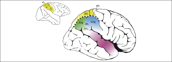
Current views of the parietal cortex have difficulty accommodating the human inferior parietal lobe (IPL) within a simple dorsal versus ventral stream dichotomy. In humans, lesions of the right IPL often lead to syndromes such as hemispatial neglect that are seemingly in accord with the proposal that this region has a crucial role in spatial processing. However, recent imaging and lesion studies have revealed that inferior parietal regions have non-spatial functions, such as in sustaining attention, detecting salient events embedded in a sequence of events and controlling attention over time. Here, we review these findings and show that spatial processes and the visual guidance of action are only part of the repertoire of parietal functions. Although sub-regions in the human superior parietal lobe and intraparietal sulcus contribute to vision-for-action and spatial functions, more inferior parietal regions have distinctly non-spatial attributes that are neither conventionally 'dorsal' nor conventionally 'ventral' in nature.


Figures

Figure 1
Figure 1
Posterior parietal cortex of macaque monkey (left) and human (right). The human posterior parietal cortex (PPC) is divided by the intraparietal sulcus (IPS) into two parts: the superior parietal lobe (SPL) and the inferior parietal lobe (IPL). The IPL consists of the angular gyrus (Ang) and supramarginal gyrus (Smg) and borders the superior temporal gyrus (purple) at a region that is often referred to as the temporoparietal junction (TPJ). In macaques, the PPC also consists of an SPL (area 5) and an IPL (areas 7a and 7b) but, according to Brodmann , the homologues of these macaque regions are all confined to the human SPL (yellow), so the IPL in humans consists of novel cortical areas. Subsequent anatomists such as Bailey and von Bonin disagreed with this scheme, considering the IPL to be similar across both species. It remains to be established whether there are new functional sub-regions within the human IPL.
Figure 2
Figure 2
Functional imaging studies. (a) Meta-analysis of activations associated with spatial shifts of attention in healthy individuals demonstrate activations in the superior parietal lobe (SPL) and intraparietal sulcus (IPS), plus dorsolateral frontal regions (different colours correspond to findings from different studies). Similar regions are also activated when participants perform spatial working memory tasks. Adapted, with permission, from Ref. , © (2002) MacMillan Publishers Ltd. (b) By contrast, regions in the inferior parietal lobe (IPL) and IPS, together with more ventral frontal regions, are activated by salient events (cyan), sustained attention (red) and non-spatial selective attention protocols (yellow) such as the attentional-blink paradigm. Adapted from Ref. .
Figure 3
Figure 3
Expansion of posterior brain regions. Human posterior brain regions in the parietotemporal regions have expanded considerably. (a) Comparison of the relative positions in macaque (left) and human (right) brains of landmark regions such as primary auditory cortex (A1) and the motion-sensitive area V5/MT reveals how the latter has shifted posteriorly and inferiorly in humans compared with its location in the depths of the superior temporal sulcus in macaque. (b) One way to map homologous regions in monkey and human brains is to compare connectivity of regions. The recent study by Rushworth et al. demonstrates connections in the human parietal cortex from the superior colliculus (connected to area LIP within the IPS of macaque), ventral premotor cortex (connected to area 7b in macaque) and the parahippocampal region (connected to area 7a in macaque). But there is a region within the human IPL (marked within the white circle) that seems not to have connections to any of these regions and might be a candidate zone for a novel cortical region within the IPL. Panel b adapted from Ref. , with permission from Oxford University Press.
Figure I
Figure I
Spatial inhomogeneity of mean lesion volume. Voxel-wise map of the rank correlation between total lesion volume and probability of damage, given a clinical and radiological diagnosis of stroke, derived from a sample of 456 patients. Only voxels affected in eight or more patients are shown. Note the strong centrifugal gradient of mean lesion size, with more peripheral (cortical) voxels being involved in larger lesions. Scale gives value of Spearman's rho.

References

    1. Mishkin M. Object vision and spatial vision: two cortical pathways. Trends Neurosci. 1983;6:414–417.
    1. Milner A.D., Goodale M.A. Oxford University Press; 1995. The Visual Brain in Action.
    1. Goodale M.A. Two distinct modes of control for object-directed action. Prog. Brain Res. 2004;144:131–144. - PubMed
    1. Milner A.D. Neglect, extinction, and the cortical streams of visual processing. In: Thier P., Karnath H-O., editors. Parietal Lobe Contributions to Orientation in 3D Space. Springer-Verlag; 1997. pp. 3–22.
    1. Vallar G., Perani D. The anatomy of unilateral neglect after right-hemisphere stroke lesions. A clinical/CT-scan correlation study in man. Neuropsychologia. 1986;24:609–622. - PubMed

Publication types