. 2007 Feb;18(2):569-80.
doi: 10.1091/mbc.e06-07-0635. Epub 2006 Nov 29.

The assembly pathway of the 19S regulatory particle of the yeast 26S proteasome

Affiliations

The assembly pathway of the 19S regulatory particle of the yeast 26S proteasome

Erika Isono et al. Mol Biol Cell. 2007 Feb.

Abstract

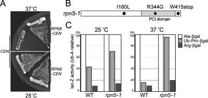
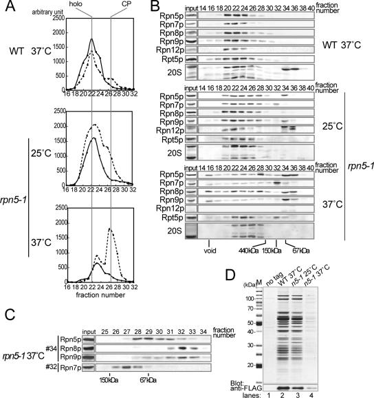
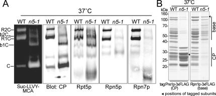
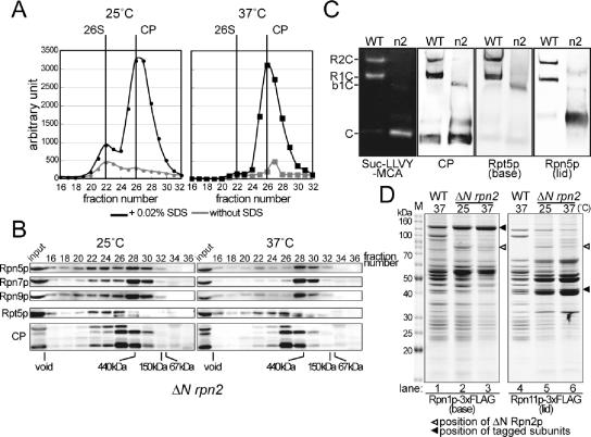
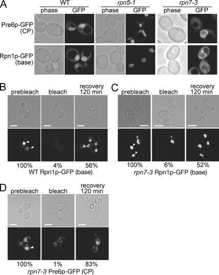
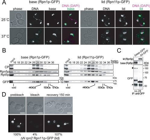
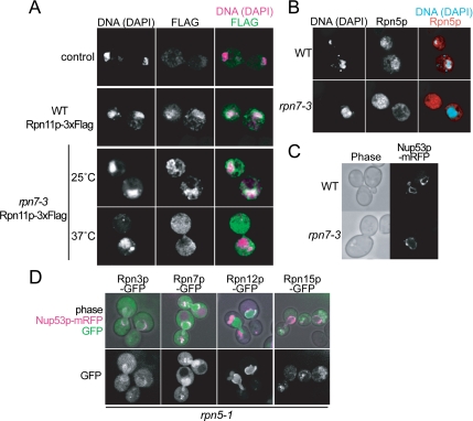
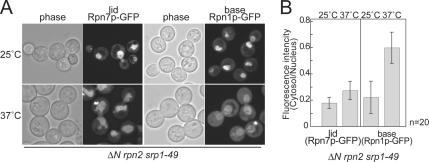
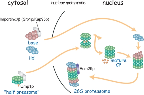
The 26S proteasome consists of the 20S proteasome (core particle) and the 19S regulatory particle made of the base and lid substructures, and it is mainly localized in the nucleus in yeast. To examine how and where this huge enzyme complex is assembled, we performed biochemical and microscopic characterization of proteasomes produced in two lid mutants, rpn5-1 and rpn7-3, and a base mutant DeltaN rpn2, of the yeast Saccharomyces cerevisiae. We found that, although lid formation was abolished in rpn5-1 mutant cells at the restrictive temperature, an apparently intact base was produced and localized in the nucleus. In contrast, in DeltaN rpn2 cells, a free lid was formed and localized in the nucleus even at the restrictive temperature. These results indicate that the modules of the 26S proteasome, namely, the core particle, base, and lid, can be formed and imported into the nucleus independently of each other. Based on these observations, we propose a model for the assembly process of the yeast 26S proteasome.


Figures

Figure 1.
Figure 1.
Characterization of the temperature-sensitive rpn5 mutant. (A) rpn5-1 (YEK100) cells carrying either a CEN vector (pRS314) or RPN5-CEN plasmid (pEK221) were streaked on YPDAU plates and photographed after incubating for 2 d at either 25 or 37°C. (B) Amino acid substitution in rpn5-1. The nucleotide sequence of the rpn5-1 ORF was determined by the dideoxychain termination method and compared with that of the wild-type RPN5 ORF. Gray, PCI domain. (C) Degradation of N-end rule pathway- and UFD pathway-substrates. Wild-type (W303-1A) and rpn5-1 (YEK100) cells were transformed with plasmids expressing an N-end rule model substrate Ub-Ala-βgal or Ub-Arg-βgal, or a UFD pathway substrate Ub-Pro-βgal. Production of the model substrates was induced by adding 2% galactose to SR-U medium. Cells were harvested after 4 h of induction at either 25 or 37°C, and steady-state levels of β-galactosidase activity were assayed. The amounts of Ub-Pro-βgal and Arg-βgal are indicated relative to that of Ala-βgal. Average of three independent experiments is shown (open, Ala-βgal; light gray, Ub-Pro-βgal; and solid, Arg-βgal).
Figure 2.
Figure 2.
Lid formation in rpn5-1 cells. Wild-type (W303-1A) and rpn5-1 (YEK100) cells were cultured for 7 h at the indicated temperature, and total cell extracts were prepared by breaking the cells by glass-beads under the existence of ATP and MgCl2. (A) Gel filtration. Peptidase activity toward the fluorogenic substrate Suc-LLVY-MCA was measured in relevant fractions (16–32). Positions of the 26S holoenzyme and the CP are indicated at the top of the graph. Solid line, without SDS; and dotted line, with 0.02% SDS. (B) Western blotting. Twenty microliters of each of the even numbered fractions was mixed with SDS-PAGE loading buffer and resolved by 12.5% SDS-PAGE, transferred to polyvinylidene difluoride membrane, and proteasome subunits were detected by Western blotting by using the indicated antibodies (Rpn5p, Rpn7p, Rpn8p, Rpn9p, and Rpn12p, lid; and Rpt5p, base) Positions of the void fraction and marker proteins (ferritin [440 kDa], aldolase [150 kDa], and bovine serum albumin [67 kDa]) are indicated at the bottom of the panels. (C) Second gel filtration. Fractions 32 and 34 in Superose 6 gel filtration of rpn5-1 extracts (37°C) in A were subsequently resolved by a Superdex 200 gel filtration column. Five hundred microliters sequential fractions were collected, and relevant fractions were subjected to Western blotting as in B. Antibodies used are indicated on the left. Positions of marker proteins (aldolase [150 kDa] and bovine serum albumin [67 kDa]) are indicated on the bottom of the panels. (D) Wild-type and rpn5-1 strains expressing RPN7-3xFLAG (YEK221 and YEK225, respectively) along with the untagged wild-type strain (W303-1A) were cultured for 7 h at 25 or 37°C as indicated, and extract was prepared from each culture. Proteasomes were affinity purified using anti-FLAG agarose. Purified products were run on a 12.5% SDS-PAGE gel, and protein bands were stained with CBB (M, marker).
Figure 3.
Figure 3.
Proteasome species in wild-type extract and rpn5-1 extract. (A) Extracts were prepared from wild-type (W303-1B) or rpn5-1 (YEK101) cells incubated for 7 h at 37°C, and extract equivalent to 50 μg of protein was resolved by nondenaturing PAGE. Proteasomes were visualized by overlaying buffer containing 0.1 mM Suc-LLVY-MCA and 0.05% SDS on the gel (far left). The gels were subsequently subjected to Western blotting by using antibodies indicated on the bottom of the panels (Rpt5p, base; and Rpn5p and Rpn7p, lid). Bands corresponding to various proteasome species are indicated on the far left of the panels (R, RP; C, CP; and b, base). (B) Affinity purification of proteasomes from CP- and base-tagged stains. YYS37 (PRE1-3xFLAG) and YYS39 (RPN1-3xFLAG), YKN6 (rpn5-1 PRE1-3xFLAG) and YKN8 (rpn5-1 RPN1-3xFLAG) cells were cultured for 7 h at 37°C and proteasomes were affinity purified from 2 mg of total proteins using anti-FLAG agarose. The purified proteasomes were resolved on a 12.5% SDS-polyacrylamide gel and stained with CBB (left, CP tagged; and right, base tagged). Bands corresponding to the tagged components are indicated with solid arrowheads. The approximate migrating positions of the base and the CP components are indicated by bars on the right side of the panel.
Figure 4.
Figure 4.
Lid formation does not depend on the binding of the lid to the base. (A) Extract of ΔN rpn2 (YAT2433) cells cultured for 6 h at 25 or 37°C was resolved on a Superose 6 column, and peptidase activity was measured as described in Figure 2A. Positions of the 26S holoenzyme and the CP are indicated at the top of the graph (black lines, with 0.02% SDS; and gray lines, without SDS). (B) Fractions were subjected to Western blotting as described in Figure 2B. Antibodies used are indicated on the left of the panel (Rpn5p, Rpn7p, and Rpn9p, lid; and Rpt5, base). Positions of the void fraction and marker proteins (ferritin [440 kDa], aldolase [150 kDa], and bovine serum albumin [67 kDa]) are indicated at the bottom of the panels. Note that all lid components examined comigrated. (C) Wild-type (W303-1A) or ΔN rpn2 (YAT2433) cells were cultured for 6 h at 37°C, and extract was prepared as described above. Extract equivalent to 50 μg of protein was resolved by nondenaturing PAGE. Proteasomes were visualized by overlaying buffer containing 0.1 mM Suc-LLVY-MCA and 0.05% SDS on the gels (far left panel). The same gels were subsequently subjected to Western blotting by using antibodies indicated on the bottom of the panels (Rpt5p, base; and Rpn5p, lid). Bands corresponding to various proteasome species are indicated on the far left of the panels (R, RP; C, CP; and b, base). (D) Affinity purification of proteasomes from base- and lid-tagged strains YYS39 (RPN1-3xFLAG), YYS40 (RPN11-3xFLAG), YEK234 (ΔN rpn2 RPN1-3xFLAG), and YAT3507 (ΔN rpn2 RPN11-3xFLAG) cells were cultured for 6 h at 25 or 37°C as indicated, and proteasomes were affinity-purified using anti-FLAG agarose. The purified proteasomes were resolved on a 12.5% SDS-polyacrylamide gel and stained with CBB (left, base tagged; and right, lid tagged). Protein bands were cut out and identified by mass spectrometry (see Supplemental Figure 2). The approximate migrating positions of base and lid components are indicated on the right of the panel (solid arrowhead, tagged component; open arrowhead, ΔN Rpn2p; and M, marker).
Figure 5.
Figure 5.
The base and the CP are localized in the nucleus in lid mutants even at the restrictive temperature. (A) Wild-type, rpn5-1 and rpn7-3 cells producing Pre6p-GFP (CP) or Rpn1p-GFP (base) instead of the authentic Pre6p and Rpn1p, respectively, were cultured for 7 h at 37°C and photographed under a confocal microscope. Strains used were PRE6-GFP (CP), wild type (YEK79), rpn5-1 (YKN18), rpn7-3 (YEK211), and RPN1-GFP (base) wild type (YEK147), rpn5-1 (YKN16), rpn7-3 (YEK213). (B–D) The base and the CP are imported into the nucleus after shift to the restrictive temperature. Rpn1p-GFP (base) producing wild-type (YEK147) and Rpn1p-GFP or Pre6p-GFP (CP) producing rpn7-3 (YEK213 and YEK211, respectively) cells were cultured for 6 h at 37°C and embedded in agarose as described in Materials and Methods. GFP signals in the nucleus (prebleach, left) were photobleached with intense laser (bleach, middle), and FRAP was observed and photographed after 120 min (recovery, right). The stage was kept at 37°C throughout the experiment. Fluorescence intensity ([/mm2] − background [/mm2]) was quantified and shown as a relative value to the prebleach intensity at the bottom of each panel. The mean value of two independent experiments is shown. Bar; 5 μm.
Figure 6.
Figure 6.
The nuclear import of the base and the lid are independent of each other. (A) ΔN rpn2 cells producing Rpn1p-GFP (base, YEK235) or Rpn11p-GFP (lid, YEK236) instead of the authentic Rpn1p or Rpn11p, respectively, were cultured for 6 h at either 25 or 37°C and photographed under a confocal microscope. DNA was stained with Hoechst 33342. (B) Total proteins were extracted from the cells cultured at 37°C as described in A and subjected to gel filtration by using a Superose 6 column. Fractions were subsequently subjected to Western blotting by using the antibodies indicated on the left of the panels. (Rpn5p; lid, Rpt5p; base) Positions of the void fraction and marker proteins (ferritin [440 kDa], aldolase [150 kDa], and bovine serum albumin [67 kDa]) are indicated at the bottom of the panels. For the GFP blot, positions of molecular mass markers are shown on the right of each of the panels. (C) Immunoprecipitation was performed against fraction 28 in B by using anti-GFP antibody and analyzed by Western blotting by using anti-Rpn5p (lid), anti-Rpt5p (base), and anti-GFP antibodies. Asterisks indicate nonspecific bands. Arrowheads indicate the GFP-fused components. (D) RPN11-GFP–expressing ΔN rpn2 (YEK256) cells were cultured for 6 h at 37°C, and FRAP experiments were performed as in Figure 5, except that the recovery was observed after 150 min. Fluorescence intensity ([/mm2] − background [/mm2]) was quantified and shown as relative values to the prebleach intensity at the bottom of each panel. The mean value of two independent experiments is shown. Arrowheads indicate the cell that was photobleached. Bar; 5 μm.
Figure 7.
Figure 7.
Localization of the partially assembled lidrpn7-3. (A) RPN11-3xFLAG (YYS40) and rpn7-3 RPN11-3xFLAG (YEK29) cells, along with untagged wild-type (W303-1A) cells, were cultured for 6 h at 25 or 37°C as indicated, and localization of lidrpn7-3 was detected by the indirect immunofluorescence method by using anti-FLAG M2 antibody. Photographs were taken under a confocal microscope. DNA was stained with DAPI. (B) Wild-type (W303-1B) and rpn7-3 (YEK6) cells were incubated for 6 h at 37°C, and localization of Rpn5p was detected as described in A by the indirect immunofluorescence method except that an anti-Rpn5p antibody was used. DNA was stained with DAPI. (C) The nuclear envelope is normal in rpn7-3 cells under the restrictive condition. Nup53p-mRFP (pEK285) was produced in wild-type (W303-1A) and rpn7-3 (YEK6) cells cultured at the same condition as described in B and photographed under a confocal microscope. (D) Localization of Rpn3p- GFP (pEK297), Rpn7p-GFP (pEK298), Rpn12p-GFP (pEK299), or Rpn15p-GFP (pEK300) in rpn5-1 (YEK100) cells. Cells were cultured for 8 h at 37°C, and GFP signals were photographed under a confocal microscope. Nup53p-mRFP was used as a marker for the nuclear envelope.
Figure 8.
Figure 8.
Nuclear localization of the base, but not the lid, is affected by srp1-49. (A) ΔN rpn2 srp1-49 cells expressing Rpn7p-GFP or Rpn1p-GFP (YEK247 and YEK258, respectively) were cultured for 8 h at either 25 or 37°C, and localization of the GFP-fused components was observed under a confocal microscope. (B) Signals of A were quantified (fluorescence intensity per area) using the IPLab software, and ratio of the nuclear and cytosolic signals is shown. Error bars represent SD (n = 20).
Figure 9.
Figure 9.
Model for the assembling process of the 26S proteasome in budding yeast. The base and the lid are made in the cytosol and are imported into the nucleus independently. On the dimerization of half-proteasomes into a mature CP, the base binds the CP. The immediate binding of the lid to the base–CP complex stabilizes the whole complex. Additional interacting proteins are bound to the 26S proteasome.

References

    1. Bachmair A., Finley D., Varshavsky A. In vivo half-life of a protein is a function of its amino-terminal residue. Science. 1986;234:179–186. - PubMed
    1. Burk D., Dawson D., Stearns T. Methods in Yeast Genetics. Cold Spring Harbor, NY: Cold Spring Harbor Laboratory Press; 2000.
    1. Cadwell R. C., Joyce G. F. Randomization of genes by PCR mutagenesis. PCR Methods Appl. 1992;2:28–33. - PubMed
    1. Campbell R. E., Tour O., Palmer A. E., Steinbach P. A., Baird G. S., Zacharias D. A., Tsien R. Y. A monomeric red fluorescent protein. Proc. Natl. Acad. Sci. USA. 2002;99:7877–7882. - PMC - PubMed
    1. Chen P., Hochstrasser M. Autocatalytic subunit processing couples active site formation in the 20S proteasome to completion of assembly. Cell. 1996;86:961–972. - PubMed

Publication types

MeSH terms

Substances

LinkOut - more resources