. 2007 Feb;18(2):627-35.
doi: 10.1091/mbc.e06-09-0839. Epub 2006 Nov 29.

Prohibitins interact genetically with Atp23, a novel processing peptidase and chaperone for the F1Fo-ATP synthase

Affiliations

Prohibitins interact genetically with Atp23, a novel processing peptidase and chaperone for the F1Fo-ATP synthase

Christof Osman et al. Mol Biol Cell. 2007 Feb.

Abstract

The generation of cellular energy depends on the coordinated assembly of nuclear and mitochondrial-encoded proteins into multisubunit respiratory chain complexes in the inner membrane of mitochondria. Here, we describe the identification of a conserved metallopeptidase present in the intermembrane space, termed Atp23, which exerts dual activities during the biogenesis of the F(1)F(O)-ATP synthase. On one hand, Atp23 serves as a processing peptidase and mediates the maturation of the mitochondrial-encoded F(O)-subunit Atp6 after its insertion into the inner membrane. On the other hand and independent of its proteolytic activity, Atp23 promotes the association of mature Atp6 with Atp9 oligomers. This assembly step is thus under the control of two substrate-specific chaperones, Atp10 and Atp23, which act on opposite sides of the inner membrane. Strikingly, both ATP10 and ATP23 were found to genetically interact with prohibitins, which build up large, ring-like assemblies with a proposed scaffolding function in the inner membrane. Our results therefore characterize not only a novel processing peptidase with chaperone activity in the mitochondrial intermembrane space but also link the function of prohibitins to the F(1)F(O)-ATP synthase complex.


Figures

Figure 1.
Figure 1.
Synthetic lethal interaction of ATP23 with PHB1 and PHB2. Tetrad dissection of 11 asci derived from a Δatp23/ATP23 Δphb1/PHB1 diploid strain (top) and a Δatp23/ATP23 Δphb2/PHB2 diploid strain (bottom). Ascospores were dissected on YPD and incubated at 30°C for 3 d. Genotypes were inferred from the distribution of the markers linked to the deletions. Double mutants are indicated with arrowheads.
Figure 2.
Figure 2.
Atp23 is an intermembrane space protein of mitochondria. (A) Mitochondria were isolated from a strain expressing Atp23-HA. Whole cell extracts (T), postmitochondrial supernatant (PMS), and isolated mitochondria (M) were analyzed by SDS-PAGE and immunoblotting using Tom40 as a mitochondrial and Bmh1 as a cytosolic marker protein. A band unspecifically cross-reacting with the Tom40-specific antiserum is marked with an asterisk. (B) Mitochondria harboring Atp23-HA were treated with trypsin (50 μg/ml) with and without hypotonic disruption (SW) of the outer membrane as described (Leonhard et al., 2000). Samples were subjected to SDS-PAGE followed by immunoblotting. Fractionation was monitored using the outer membrane protein Tom70, the intermembrane space protein Yme1 and the matrix-localized protein Mge1 as controls. (C) Isolated mitochondria were treated with sodium carbonate (pH 11.5, T) and subjected to ultracentrifugation to obtain soluble (S) and insoluble (P) fractions, which were analyzed by SDS-PAGE and immunoblotting. The integral inner membrane protein Yme1 and the soluble intermembrane space protein cytochrome b2 (Cyb2) were used as controls.
Figure 3.
Figure 3.
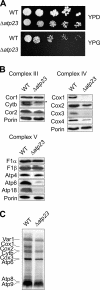
Atp23 is essential for respiratory growth. (A) Serial dilutions of wild-type (WT) and Δatp23 cells were plated on YPD and YPG plates. Strains were grown at 30°C. (B) Steady state levels of various subunits of the respiratory chain were examined by immunoblotting of mitochondria (50 μg) derived from Δatp23 and wild-type (WT) cells. A band unspecifically cross-reacting with cytochrome b (Cytb)-specific antiserum is marked with an asterisk. (C) Synthesis of mitochondrial-encoded proteins in wild-type (WT) and Δatp23 mitochondria. Mitochondrial translation products were synthesized in the presence of [35S]methionine. Mitochondrial proteins were separated by SDS-PAGE and analyzed by autoradiography. The efficiency of Cox1 labeling varied in different experiments (data not shown).
Figure 4.
Figure 4.
Dual activity of Atp23 within mitochondria. (A) Accumulation of uncleaved Atp6 in mitochondria harboring mutant Atp23. Mitochondrial protein synthesis was carried out in the presence of [35S]methionine in mitochondria isolated from wild-type (WT) cells and from Δatp23 cells that expressed Atp23 variants carrying mutations in the consensus metal binding site (Atp23H167A, Atp23E168Q, Atp23H171A) when indicated. Mitochondrial proteins were analyzed by SDS-PAGE and Western blotting. An autoradiograph of the membrane is shown in the top panel (35S), an immunoblot analysis with Atp6- and porin-specific antisera in the middle and bottom panels. p, precursor form; m, mature form. (B) Binding of newly synthesized Atp6 to Atp23-HA. After labeling of mitochondrial translation products with [35S]methionine in wild-type (WT) mitochondria and mitochondria harboring Atp23-HA (HA), mitochondria were lysed with digitonin. Mitochondrial extracts (load, 5% of total) were subjected to coimmunoprecipitation with HA-specific antiserum. The precipitates (prec) and unbound material (unb, 10% of total) were analyzed by SDS-PAGE and autoradiography. The top panel shows the autoradiograph including all mitochondrial translation products. In the bottom panel, a part of an autoradiograph of a high-resolution gel is shown including Cox3, Atp6 precursor (p), and mature Atp6 (m). (C) Respiratory growth of Δatp23 cells expressing mutant Atp23. Atp23 variants containing mutations in the consensus metal-binding site (Atp23H167A, Atp23E168Q, Atp23H171A) were expressed in Δatp23 cells. Fivefold serial dilutions of wild-type (WT) and mutant cells were plated on YPD and YPG and incubated for 2 d at 30°C. (D) Multiple sequence alignment of Atp23 and homologues using AlignX. Sequences surrounding the consensus metal-binding sites are shown. Sequence identities between whole proteins according to Blastp are indicated. Sc, S. cerevisiae Atp23 (Ynr020c), Cg, Candida glabrata CAG62785; Nc, Neurospora crassa XP322193; At, Arabidopsis thaliana Ku70-binding family protein NP_566205; Dm, Drosophila melanogaster CG5131-PA; Hs, Homo sapiens KUB3; and Xl, Xenopus laevis KUB3-homologue. Identical amino acids are shown in black, conserved residues in dark gray, and similar residues in light gray.
Figure 5.
Figure 5.
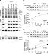
Atp23 is required for the assembly of the FO-particle of the F1FO-ATP synthase independent of its proteolytic activity. (A) Wild-type and Δatp23 mitochondria (100 μg), which contain Atp23 variants carrying mutations in the consensus metal binding site (Atp23H167A, Atp23E168Q, Atp23H171A) when indicated, were solubilized in digitonin (1.8%) and analyzed by BN-PAGE followed by Coomassie staining (top panel) or immunoblotting (lower three panels) using polyclonal antisera directed against Atp6, the α-subunit of the F1-particle (F1α), or subunit 2 of cytochrome c oxidase (Cox2). The position of supercomplexes containing complex III and IV (III2IV2; III2IV1) and of monomeric (Vmon) and dimeric (Vdim) F1FO-ATP synthase complexes are indicated. (B and C) Assembly of newly synthesized, mitochondrial-encoded FO-subunits Atp6, Atp8, and Atp9 into F1FO-ATP synthase complexes. Cells were grown in the presence of chloramphenicol before mitochondria were isolated. Mitochondrial-encoded proteins were labeled with [35S]methionine and, after inhibition of translation, mitochondria were further incubated at 30°C for the time indicated to allow the assembly of newly synthesized subunits. Mitochondria (150 μg) were solubilized in 1.6% (vol/vol) Triton X-100 and subjected to BN-PAGE. Only monomeric F1FO-ATP synthase is detectable under these conditions (Arnold et al., 1998). Coomassie stained gels (Com) and autoradiographs (35S) are shown in the top panel. Incorporated Radioactivity incorporated into monomeric ATP synthase was quantified by phosphoimaging and corrected for different labeling efficiencies in various mitochondria. (B) Analysis of F1FO-ATP synthase assembly in wild-type (WT) and Δatp23 mitochondria. (C) Analysis of F1FO-ATP synthase assembly in Δatp23 mitochondria harboring Atp23 (ATP23) or the mutant variant Atp23E168Q (E168Q).
Figure 6.
Figure 6.
Genetic interaction of PHB1 with ATP23 and ATP10 controlling Atp6 assembly into the FO-particle. (A) Proteolytically inactive Atp23 allows growth of Δphb1Δatp23 cells. Tetrads derived from diploid Δphb1/PHB1 Δatp23 cells expressing Atp23 or its proteolytically inactive variants Atp23H167A, Atp23E168Q, Atp23H171A were dissected. Ascospores carrying deletion of both PHB1 and ATP23 and expressing Atp23 or variants thereof were isolated and examined for growth on YPD and YPG at 30°C. (B) Synthetic lethal interaction of PHB1 with ATP10. The genetic interaction of ATP10 with PHB1 was confirmed by tetrad analysis as described in Figure 1 for ATP23. Similarly, a synthetic lethal interaction was observed between ATP10 and PHB2 (data not shown). (C) Impaired assembly of F1FO-ATP synthase complexes in Δatp10 and Δatp23 mitochondria. The assembly of the F1FO-ATP synthase was analyzed by BN-PAGE after solubilization of mitochondrial membranes with digitonin as described in Figure 5A. Assembly intermediates detected in Δatp23 and, to a lower extent, in Δatp10 mitochondria are marked with asterisks.
Figure 7.
Figure 7.
Model of Atp6 assembly into F1FO-ATP synthase complexes. See text for details. The translocase mediating membrane insertion of Atp6 has not been identified. 10, Atp10; 23, Atp23; 9, Atp9; mAAA, m-AAA protease.

References

    1. Ackerman S. H., Tzagoloff A. Identification of two nuclear genes (ATP11, ATP12) required for assembly of the yeast F1-ATPase. Proc. Natl. Acad. Sci. USA. 1990;87:4986–4990. - PMC - PubMed
    1. Ackerman S. H., Tzagoloff A. Function, structure, and biogenesis of mitochondrial ATP synthase. Prog. Nucleic Acid Res. Mol. Biol. 2005;80:95–133. - PubMed
    1. Ackermann S. H., Tzagoloff A. ATP10, a yeast nuclear gene required for the assembly of the mitochondrial F1-F0 complex. J. Biol. Chem. 1990;265:9952–9959. - PubMed
    1. Arlt H., Steglich G., Perryman R., Guiard B., Neupert W., Langer T. The formation of respiratory chain complexes in mitochondria is under the proteolytic control of the m-AAA protease. EMBO J. 1998;17:4837–4847. - PMC - PubMed
    1. Arnold I., Pfeiffer K., Neupert W., Stuart R. A., Schägger H. Yeast mitochondrial F1FO-ATP synthase exists as a dimer: identification of three dimer-specific subunits. EMBO J. 1998;17:7170–7178. - PMC - PubMed

Publication types

MeSH terms