

doi: 10.1128/JVI.01781-06.
Epub 2006 Nov 29.
Evidence for the existence of the loop E motif of Potato spindle tuber viroid in vivo
Affiliations
- PMID: 17135317
- PMCID: PMC1797592
- DOI: 10.1128/JVI.01781-06
Item in Clipboard
Evidence for the existence of the loop E motif of Potato spindle tuber viroid in vivo
J Virol.
2007 Feb.
Abstract
RNA motifs comprising nucleotides that interact through non-Watson-Crick base pairing play critical roles in RNA functions, often by serving as the sites for RNA-RNA, RNA-protein, or RNA small ligand interactions. The structures of viral and viroid RNA motifs are studied commonly by in vitro, computational, and mutagenesis approaches. Demonstration of the in vivo existence of a motif will help establish its biological significance and promote mechanistic studies on its functions. By using UV cross-linking and primer extension, we have obtained direct evidence for the in vivo existence of the loop E motif of Potato spindle tuber viroid. We present our findings and discuss their biological implications.
Figures
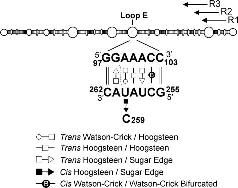
Secondary structure of PSTVd (upper panel) (14) and non-Watson-Crick base pairs in loop E (lower panel) (37). The symbols that denote each of the specific base edge-edge interactions are defined below the structure (for details, see references and 37). The dashed line indicates G98 and U260, which can be cross-linked by UV irradiation. R1, R2, and R3 indicate the positions of the primers used in the primer extension experiments.
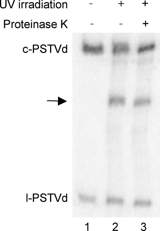
RNA gel blot showing the appearance of a UV-cross-linked product (arrow) of PSTVd RNAs from infected tomato leaves. Treatment with proteinase K (0.5 units for 30 min; Invitrogen) indicates that this cross-linked product is not attributed to protein binding. c-PSTVd and l-PSTVd denote circular and linear PSTVd RNAs, respectively. For gel blotting, total RNAs were extracted from infected tomato leaves that were untreated or irradiated with UV for 80 min (10 J/cm2) by using the RNeasy plant mini kit (QIAGEN, Valencia, CA). RNA samples were run on 5% polyacrylamide-8 M urea gels, transferred to Hybond-XL nylon membranes (Amersham Biosciences, Piscataway, NJ) using a vacuum blotting system (Amersham), and immobilized by UV cross-linking. After overnight hybridization with [α-32P]UTP-labeled riboprobes at 65°C in ULTRAhyb reagent (Ambion, Austin, TX), the membranes were washed twice at 65°C in 2× SSC-0.1% sodium dodecyl sulfate (1× SSC is 0.15 M NaCl plus 0.015 M sodium citrate) and 0.2× SSC-0.1% sodium dodecyl sulfate and exposed to a Storage phosphor screen (Kodak, Rochester, NY).
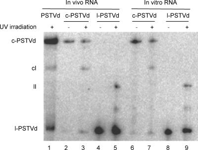
RNA gel blot showing the origin of the in vivo UV-cross-linked product predominantly from the circular PSTVd RNAs. Total RNAs were extracted and run on 5% polyacrylamide-8 M urea gels as described in the legend for Fig. 2. The in vivo circular PSTVd (c-PSTVd) and linear PSTVd (l-PSTVd) RNAs were gel purified and irradiated with UV for 5 min in EXL buffer (600 mM NaCl, 4 M urea, 1 mM cacodylate, 0.1 mM EDTA) as described by Zhong et al. (37). In vitro transcripts of PSTVd were obtained by transcribing HindIII-linearized pRZ6-2 template (17) by utilizing a T7 MAXIscript kit (Ambion). Unit-length l-PSTVd RNAs were gel purified from the 5% polyacrylamide-8 M urea gel. Incubation of the l-PSTVd transcripts in wheat germ extract gave rise to the circular PSTVd RNAs, which were run on a 5% polyacrylamide-8 M urea gel followed by gel purification. Both c-PSTVd and l-PSTVd in vitro transcripts were subjected to UV irradiation for 5 min in the EXL buffer. Approximately 100 to 200 ng of RNA was loaded for each lane.
Primer extension mapping of an in vivo cross-linking site to G98 of loop E, using three different primers as indicated (R1, R2, and R3). Lane c, cDNAs from reverse transcription of the circular PSTVd RNA template purified from infected tomato leaves; lane cI, cDNAs from reverse transcription of the UV cross-linked circular PSTVd RNA template purified from infected leaves. The reverse transcription was performed by following the protocols of Baumstark et al. (3) with modifications, using Invitrogen Superscript III RNase H- reverse transcriptase and 32P-labeled primers for 40 min at 52°C. Lanes U to A, sequencing ladders generated by PCR with pRZ6-2 template and 32P-labeled primers in the presence of ddATP, ddCTP, ddGTP, and ddUTP by utilizing the Thermo Sequenase cycle sequencing kit (USB, Cleveland, OH). All samples were run on an 8% polyacrylamide-8 M urea sequencing gel. The nucleotide sequence of the (+)-PSTVd is given on the left, and the arrows point to the bands corresponding to reverse transcription termination at A99.
Similar articles
-
Secondary structure probing of potato spindle tuber viroid (PSTVd) and sequence comparison with other small pathogenic RNA replicons provides evidence for central non-canonical base-pairs, large A-rich loops, and a terminal branch.J Mol Biol. 1996 Oct 11;262(5):652-70. doi: 10.1006/jmbi.1996.0543. J Mol Biol. 1996. PMID: 8876645
-
Existence in vivo of the loop E motif in potato spindle tuber viroid RNA.Arch Virol. 2007;152(7):1389-93. doi: 10.1007/s00705-007-0952-y. Epub 2007 Mar 19. Arch Virol. 2007. PMID: 17370107
-
Tertiary structural and functional analyses of a viroid RNA motif by isostericity matrix and mutagenesis reveal its essential role in replication.J Virol. 2006 Sep;80(17):8566-81. doi: 10.1128/JVI.00837-06. J Virol. 2006. PMID: 16912306 Free PMC article.
-
Viroid: a useful model for studying the basic principles of infection and RNA biology.Mol Plant Microbe Interact. 2007 Jan;20(1):7-20. doi: 10.1094/MPMI-20-0007. Mol Plant Microbe Interact. 2007. PMID: 17249418 Review.
-
Discovering viroids--a personal perspective.Nat Rev Microbiol. 2003 Oct;1(1):75-80. doi: 10.1038/nrmicro736. Nat Rev Microbiol. 2003. PMID: 15040183 Review.
Cited by
-
Viroid replication: rolling-circles, enzymes and ribozymes.Viruses. 2009 Sep;1(2):317-34. doi: 10.3390/v1020317. Epub 2009 Sep 14. Viruses. 2009. PMID: 21994552 Free PMC article.
-
Structural differences within the loop E motif imply alternative mechanisms of viroid processing.RNA. 2007 Jun;13(6):824-34. doi: 10.1261/rna.452307. Epub 2007 Apr 16. RNA. 2007. PMID: 17438124 Free PMC article.
-
Function and evolution of a MicroRNA that regulates a Ca2+-ATPase and triggers the formation of phased small interfering RNAs in tomato reproductive growth.Plant Cell. 2011 Sep;23(9):3185-203. doi: 10.1105/tpc.111.088013. Epub 2011 Sep 13. Plant Cell. 2011. PMID: 21917547 Free PMC article.
-
A genomic map of viroid RNA motifs critical for replication and systemic trafficking.Plant Cell. 2008 Jan;20(1):35-47. doi: 10.1105/tpc.107.056606. Epub 2008 Jan 4. Plant Cell. 2008. PMID: 18178767 Free PMC article.
-
Tertiary structure and function of an RNA motif required for plant vascular entry to initiate systemic trafficking.EMBO J. 2007 Aug 22;26(16):3836-46. doi: 10.1038/sj.emboj.7601812. Epub 2007 Jul 26. EMBO J. 2007. PMID: 17660743 Free PMC article.
References
-
- Branch, A. D., B. J. Benenfeld, B. M. Baroudy, F. V. Wells, J. L. Gerin, and H. D. Robertson. 1989. An ultraviolet-sensitive RNA structural element in a viroid-like domain of the hepatitis delta virus. Science 243:649-652. - PubMed
Publication types
MeSH terms
Substances
LinkOut - more resources
Full Text Sources
