. 2007 Feb;81(4):1641-9.
doi: 10.1128/JVI.01671-06. Epub 2006 Nov 29.

Regulation of hepadnavirus reverse transcription by dynamic nucleocapsid phosphorylation

Affiliations

Regulation of hepadnavirus reverse transcription by dynamic nucleocapsid phosphorylation

Suresh H Basagoudanavar et al. J Virol. 2007 Feb.

Abstract

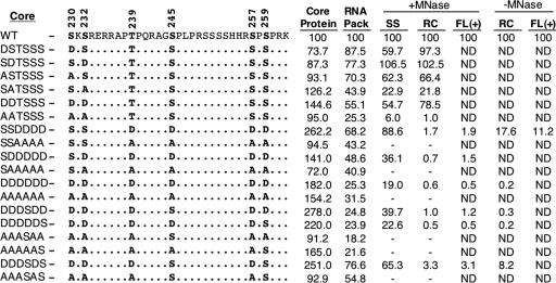
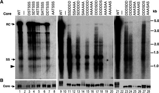
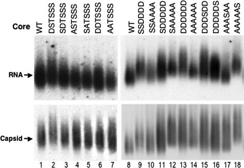
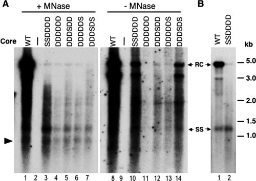
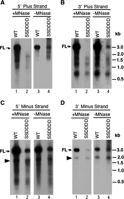
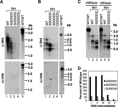
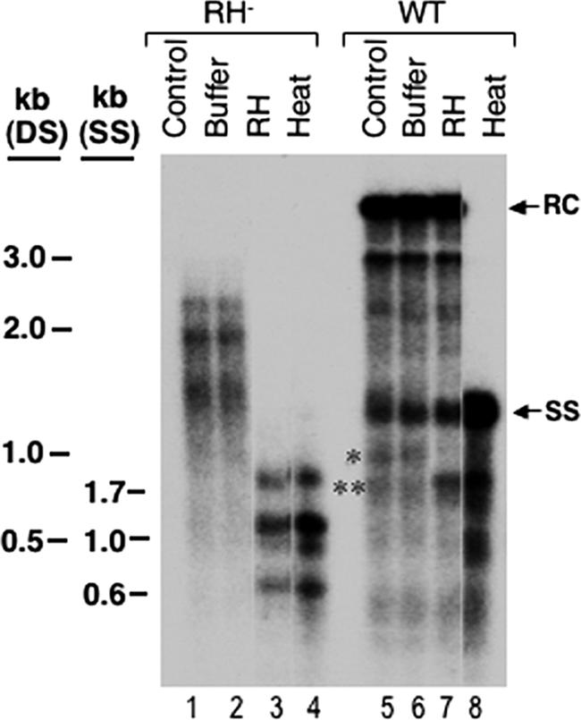
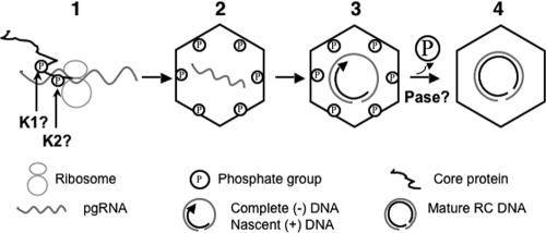
Reverse transcription, an essential step in the life cycle of all retroelements, is a complex, multistep process whose regulation is not yet clearly understood. We have recently shown that reverse transcription in the pararetrovirus duck hepatitis B virus is associated with complete dephosphorylation of the viral core protein, which forms the nucleocapsid wherein reverse transcription takes place. Here we present a genetic study of the role of this dynamic nucleocapsid phosphorylation in regulating viral reverse transcription. Detailed analyses of the reverse transcription products synthesized within nucleocapsids composed of core phosphorylation site mutants revealed that alanine substitutions, mimicking the nonphosphorylated state, completely blocked reverse transcription at a very early stage. In contrast, aspartate substitutions, mimicking the phosphorylated state, allowed complete first-strand DNA synthesis but were severely defective in accumulating mature double-stranded DNA. The latter defect was due to a combination of mutant nucleocapsid instability during maturation and a block in mature second-strand DNA synthesis. Thus, the reversible phosphorylation of the nucleocapsids regulates the ordered progression of reverse transcription.


Figures

FIG. 1.
FIG. 1.
Summary of DHBV core phosphorylation mutants. The sequences of the C-terminal 33 amino acids of the WT and various mutants are shown. The S or T residues at the six known phosphorylation sites (shown in bold) were changed to either A or D, as indicated. The mutants are designated on the left. The average levels in the various core mutants, compared to those in the WT (set to 100), of core protein expression (as measured by SDS-PAGE and Western blot analysis), pgRNA packaging (RNA pack) (as measured by the native agarose gel electrophoresis assay), SS DNA (extracted with micrococcal nuclease [MNase] digestion), RC DNA (extracted with or without exogenous nuclease digestion), and full-length plus-strand DNA [FL (+), extracted with or without nuclease digestion and measured using denatured core DNA], are indicated. ND, not determined; −, levels of DNA were too low to be quantified reliably.
FIG. 2.
FIG. 2.
Effects of core substitutions on protein expression and DNA synthesis. LMH cells were cotransfected with pCMV-DHBV/C, which expresses a core-defective DHBV genome, together with the indicated core (WT or mutant) expression plasmid. (A) Replicative viral DNA intermediates were isolated (with nuclease digestion [see Materials and Methods for details]) from the cytoplasmic NCs and analyzed by Southern hybridization using a radiolabeled DHBV DNA probe. (B) Core protein expression was analyzed by SDS-PAGE and Western blotting with a core-specific antibody. RC, relaxed circular DNA; SS, full-length single-stranded DNA. The arrowhead indicates the minus-strand DNA (ca. 2.0 kb) reverse transcribed from an internally deleted pgRNA. See the text for details. The marker DNA sizes (kb) are indicated.
FIG. 3.
FIG. 3.
Effects of core mutations on pgRNA packaging. LMH cells were cotransfected as described in the legend to Fig. 2, except that the core-defective DHBV genome also harbored a mutant polymerase defective in DNA synthesis but competent for pgRNA packaging (DHBV/C/YMHA). Cytoplasmic capsids were analyzed by native agarose gel electrophoresis and transferred to a nitrocellulose membrane. A radiolabeled plus-strand-specific riboprobe was used to detect the packaged pgRNA (top), and an anticore antibody was used to detect the capsids on the same membrane (bottom).
FIG. 4.
FIG. 4.
Comparative analysis of viral core DNA isolated with or without nuclease digestion. (A) LMH cells were transfected as described in the legend to Fig. 2. Core DNA was isolated either following micrococcal nuclease (MNase) treatment of cytoplasmic lysates (lanes 1 to 7) or by the SDS-KCl precipitation method without nuclease treatment (lanes 8 to 14) (see Materials and Methods for details) and analyzed by Southern blot hybridization using a radiolabeled DHBV DNA probe. (B) LMH cells were transfected as described in the legend to Fig. 2, except that the complementing construct used was pcDNA-Dcore (rather than pCMVDHBVΔXM) expressing either the WT or the SSDDDD mutant core protein. RC, relaxed circular DNA; SS, full-length single-stranded DNA. The arrowhead in panel A indicates the internally deleted minus-strand DNA, as described in the legend to Fig. 2.
FIG. 5.
FIG. 5.
Analysis of denatured core DNA. Viral core DNA was isolated from transfected LMH cells, with or without micrococcal nuclease (MNase) digestion, and heat denatured prior to Southern blot analysis. Radiolabeled specific riboprobes hybridizing to the plus (A [5′ end, nt 2651 to 415] and B [3′ end, nt 2156 to 2375])- or minus (C [5′ end, nt 2156 to 2531] and D [3′ end, nt 2651 to 3021])-strand DNA were used to detect the different regions and strands of viral DNA. The DNA marker sizes (kb) (heat denatured) are indicated. FL, full-length plus (A and B)- or minus (C and D)-strand DNA. The arrowheads in panels C and D indicate the internally deleted minus-strand DNA described in the legend to Fig. 2.
FIG. 6.
FIG. 6.
Effects of core mutations on minus-strand DNA synthesis in RNase H-defective NCs. Viral core DNA was isolated from transfected LMH cells as described in the legend to Fig. 2, except that the core-defective DHBV genome also harbored a mutant polymerase defective in RNase H activity (DHBV/C/RH). DNAs were extracted with (A, B, and C, lanes 1 to 3) or without (C, lanes 4 to 6) exogenous micrococcal nuclease (MNase) digestion and analyzed by Southern hybridization, with (B and C) or without (A) prior heat denaturation. Minus-strand DNA was detected using a radiolabeled riboprobe specific for the 5′ end of the minus strand (as in Fig. 5C), and plus-strand DNA was detected using a full-length riboprobe. The marker DNA sizes (kb), either DS (A) or denatured SS (B and C), are indicated. The numerals I through V in panel B refer to minus-strand DNA intermediates with increasing lengths. Lanes 5 in panels A and B and lanes 1 and 4 in panel C show the DNA synthesized by the WT polymerase in the context of the WT NC (WT/WT), as a control. (D) Relative amounts of minus-strand intermediates, designated I through V as in panel B, are expressed as percentages of those in the WT. Note that all comparisons in panel D were made among NCs that shared the same RNase H mutation.
FIG. 7.
FIG. 7.
Analysis of RNA-DNA hybrids in RNase H-defective NCs. Viral core DNA isolated from the RNase H-defective mutant (RH) or the WT was treated with RNase H (RH) (lanes 3 and 7) or buffer alone (lanes 2 and 6) or heat denatured at 95°C (lanes 4 and 8) and then analyzed by Southern blot hybridization using a riboprobe specific for the 5′ end of the minus-strand DNA (as in Fig. 5C). RC, relaxed circular DNA; SS, full-length single-stranded DNA. The marker DNA (SS, heat-denatured, single-stranded DNA; DS, double-stranded DNA) sizes are indicated. In addition to the predicted RNA-DNA hybrid present in the RH mutant (lanes 1 and 2), an apparent RNA-DNA hybrid (*) was also present in the WT core DNA (lanes 5 and 6) and was converted to a faster-migrating short minus-strand DNA (**) after RNase H digestion (lane 7) or heating (lane 8).
FIG. 8.
FIG. 8.
Model of sequential core phosphorylation and dephosphorylation regulating hepadnavirus reverse transcription. (1) Core phosphorylation during or after translation; (2) NC assembly and pgRNA packaging; (3) complete minus-strand DNA synthesis and initial stage of plus-strand DNA synthesis; (4) core dephosphorylation followed by plus-strand DNA maturation. K1 and K2, unknown cellular kinases; Pase, unknown cellular phosphatase. See the text for details.

Similar articles

Cited by

References

    1. Birnbaum, F., and M. Nassal. 1990. Hepatitis B virus nucleocapsid assembly: primary structure requirements in the core protein. J. Virol. 64:3319-3330. - PMC - PubMed
    1. Bottcher, B., S. A. Wynne, and R. A. Crowther. 1997. Determination of the fold of the core protein of hepatitis B virus by electron cryomicroscopy. Nature 386:88-91. - PubMed
    1. Bowzard, J. B., J. W. Wills, and R. C. Craven. 2001. Second-site suppressors of Rous sarcoma virus Ca mutations: evidence for interdomain interactions. J. Virol. 75:6850-6856. - PMC - PubMed
    1. Chang, L. J., R. C. Hirsch, D. Ganem, and H. E. Varmus. 1990. Effects of insertional and point mutations on the functions of the duck hepatitis B virus polymerase. J. Virol. 64:5553-5558. - PMC - PubMed
    1. Chen, Y., and P. L. Marion. 1996. Amino acids essential for RNase H activity of hepadnaviruses are also required for efficient elongation of minus-strand viral DNA. J. Virol. 70:6151-6156. - PMC - PubMed

Publication types

Substances

LinkOut - more resources