Comparative Study
. 2006 Nov 29;26(48):12497-511.
doi: 10.1523/JNEUROSCI.3719-06.2006.

Transplantation of human neural stem cells exerts neuroprotection in a rat model of Parkinson's disease

Affiliations
Comparative Study

Transplantation of human neural stem cells exerts neuroprotection in a rat model of Parkinson's disease

Takao Yasuhara et al. J Neurosci. .

Abstract

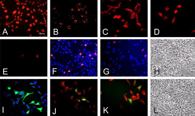
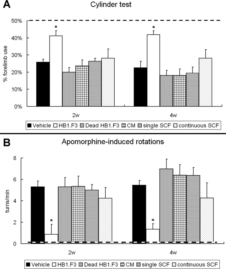
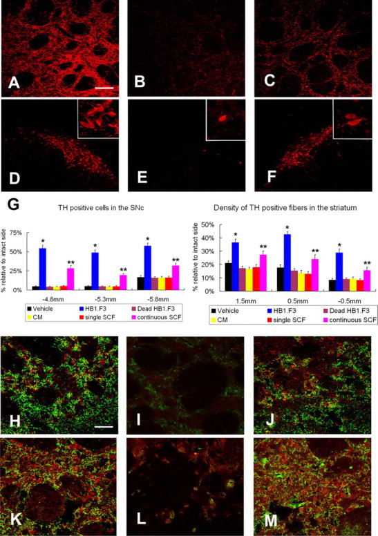
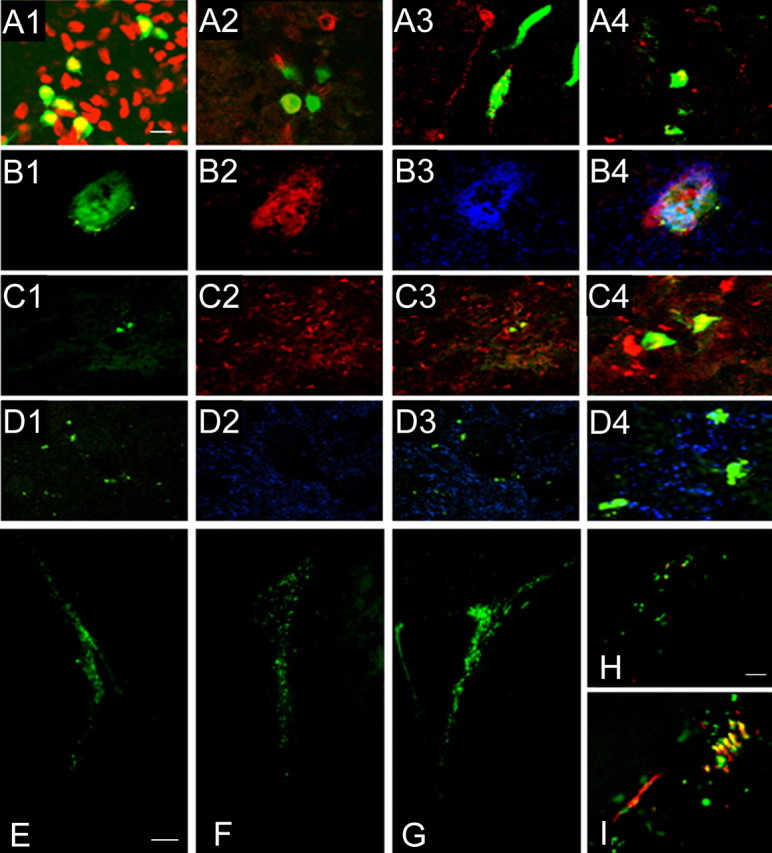
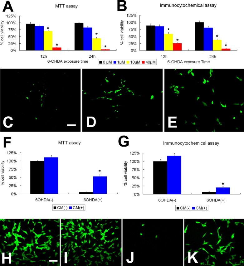
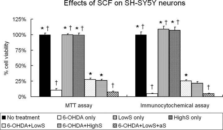
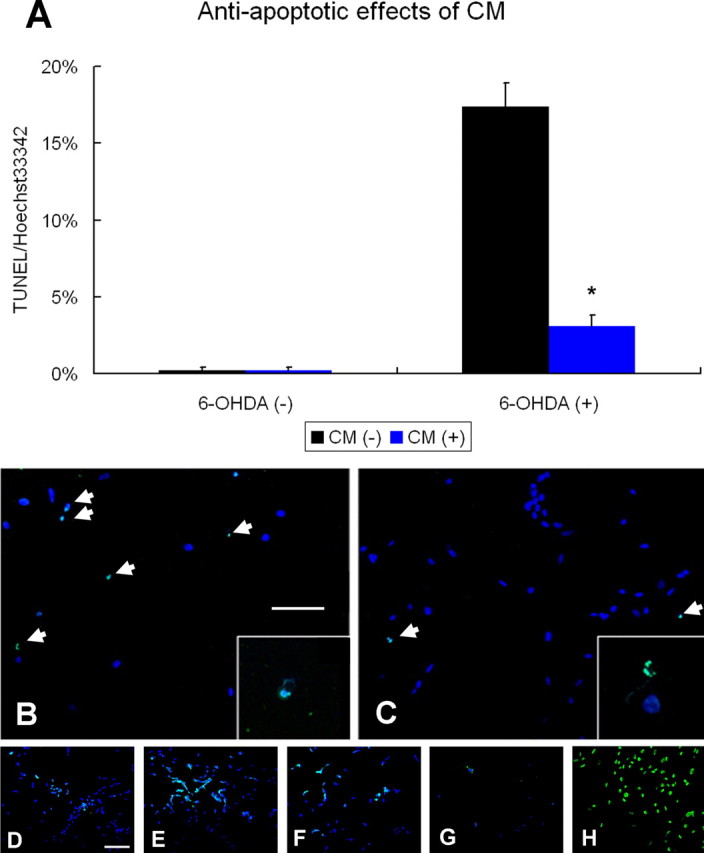
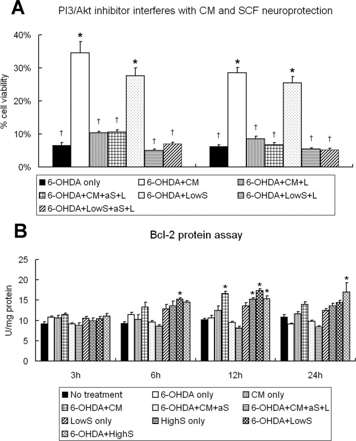
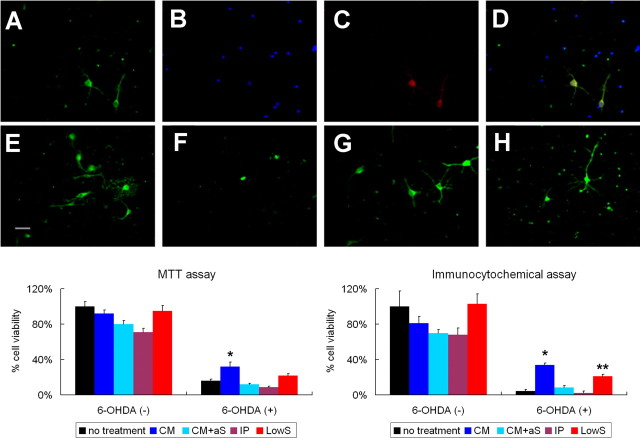
Neural stem cells (NSCs) possess high potencies of self-renewal and neuronal differentiation. We explored here whether transplantation of human NSCs cloned by v-myc gene transfer, HB1.F3 cells, is a feasible therapeutic option for Parkinson's disease. In vivo, green fluorescent protein-labeled HB1.F3 cells (200,000 viable cells in 3 microl of PBS) when stereotaxically transplanted (same-day lesion-transplant paradigm) into the 6-hydroxydopamine-lesioned striatum of rats significantly ameliorated parkinsonian behavioral symptoms compared with controls (vehicle, single bolus, or continuous minipump infusion of trophic factor, or killed cell grafts). Such graft-derived functional effects were accompanied by preservation of tyrosine hydroxylase (TH) immunoreactivity along the nigrostriatal pathway. Grafted HB1.F3 cells survived in the lesioned brain with some labeled with neuronal marker mitogen-activated protein 2 and decorated with synaptophysin-positive terminals. Furthermore, endogenous neurogenesis was activated in the subventricular zone of transplanted rats. To further explore the neuroprotective mechanisms underlying HB1.F3 cell transplantation, we performed cell culture studies and found that a modest number of HB1.F3 cells were TH and dopamine and cAMP-regulated phosphoprotein 32 positive, although most cells were nestin positive, suggesting a mixed population of mature and immature cells. Administration of the HB1.F3 supernatant to human derived dopaminergic SH-SY5Y cells and fetal rat ventral mesencephalic dopaminergic neurons protected against 6-hydroxydopamine neurotoxicity by suppressing apoptosis through Bcl-2 upregulation, which was blocked by anti-stem cell factor antibody alone, the phosphatidylinositol 3-kinase/Akt inhibitor LY294002 [2-(4-morpholinyl)-8-phenyl-1(4H)-benzopyran-4-one] alone, or a combination of both. These results suggest that HB1.F3 cell transplantation exerts neuroprotective effects against dopaminergic depletion in vitro and in vivo because of trophic factor secretion and neuronal differentiation.


Figures

Figure 1.
Figure 1.
Phenotypic characterization of HB1.F3 cells and GFP–HB1.F3 cells in vitro. Immunocytochemical evaluation of HB1.F3 cells revealed that several cells were positive for nestin, human nuclei, HuD, and β-tubulin III antibody, some stained for MAP2, and some rarely labeled with NeuN. Some cells were DARPP32 and TH positive, thus indicating that the cells display the characteristics of striatal neurons, although their morphological immature appearance suggests an early progenitor cell type. Parallel immunocytochemical evaluations of GFP–HB1.F3 cells revealed the same expression of phenotypic markers and morphology found in non-viral labeled HB1.F3 cells. A, Nestin; B, human nuclei; C, HuD; D, MAP2; E, NeuN; F, DARPP32 (red) and Hoechst33342 (blue); G, TH (red) and Hoechst33342 (blue); H, phase-contrast image of HB1.F3 cells; I, GFP–HB1.F3 cells (blue, Hoechst33342); J, β-tubulin III (red); K, MAP2 (red); L, phase-contrast image of GFP–HB1.F3 cells. Scale bar, 30 μm.
Figure 2.
Figure 2.
HB1.F3 cell grafts ameliorate forelimb akinesia and apomorphine-induced rotations. A, Cylinder test revealed improved forelimb akinesia in lesioned rats that received HB1.F3 cell grafts compared with control rats, except for rats receiving continuous SCF infusion at 2 and 4 weeks after transplantation. Dashed line indicates performance of control (i.e., naive non-6-OHDA-lesioned age-matched rats). Data are shown as mean ± SE ratio of the affected forelimb use. *p < 0.05 versus control rats except for rats receiving continuous SCF infusion. B, The number of apomorphine-induced rotations in lesioned rats that received HB1.F3 cell grafts significantly decreased compared with control rats, except for rats receiving continuous SCF infusion at 2 and 4 weeks. Dashed line indicates performance of control, nonlesioned rats. Data are shown as mean ± SE rotations per minute. *p <0.05 versus control rats, except for rats receiving continuous SCF infusion.
Figure 3.
Figure 3.
HB1.F3 cell grafts preserve TH-positive fibers in the striatum and neurons in the SNc and normalize c-kit/TrkB expression in the striatum. A–F, TH-positive fibers in the striatum and neurons in the SNc of rats that received HB1.F3 cell grafts were markedly preserved (C, F) compared with vehicle-treated rats (B, E). Intact striatum (A) and intact SNc (D) show normal TH-positive fibers and neurons, respectively. Insets are representative higher magnifications of TH-positive cells in D–F. Scale bar: A–C (captured by confocal microscopy), 50 μm; D–F, 100 μm; insets in D–F, 10 μm. G, Left, TH-positive fibers at three different coronal levels in the striatum were analyzed with a computerized image analysis system. Lesioned rats transplanted with HB1.F3 cells exhibited significantly more TH-positive fibers than all other lesioned rats. Rats with continuous SCF infusion also demonstrated significantly more TH-positive neurons than vehicle-treated, dead HB1.F3-transplanted, CM and single SCF-injected lesioned rats. Data are shown as mean ± SE values expressed as percentages relative to the intact side. *p < 0.05 versus all other rats; **p < 0.05 versus vehicle-treated, dead HB1.F3-transplanted, CM and single SCF-injected lesioned rats. G, Right, TH-positive fibers at three different levels in the striatum were analyzed with a computerized image analysis system. Lesioned rats transplanted with HB1.F3 cells demonstrated significantly more TH-positive fibers than all other lesioned rats. Rats with continuous SCF infusion also demonstrated significantly more TH-positive fibers than vehicle-treated, dead HB1.F3-transplanted, CM and single SCF-injected lesioned rats. Data are shown as mean values ± SE expressed as percentages relative to the intact side. *p < 0.05 versus all other rats; **p < 0.05 versus vehicle-treated, dead HB1.F3-transplanted, CM and single SCF-injected lesioned rats. H–M, The expression of the SCF receptor c-kit and the BDNF receptor TrkB was confirmed immunohistochemically in the intact and transplanted striatum by confocal microscopy. Yellowish fibers indicate colabeling of TH-positive fibers (green) with one of the receptors (red). The expression of both receptors in the striatum of vehicle-treated lesioned rats was difficult to identify because of severely reduced TH-positive fibers. H–J, TH (green) and c-kit (red). K–M, TH (green) and TrkB (red). H and K correspond to intact striatum, I and L represent vehicle-treated lesioned striatum, and J and M are from HB1.F3-transplanted lesioned striatum. Scale bar. 30 μm
Figure 4.
Figure 4.
HB1.F3 cell grafts survive in the lesioned striatum and enhance neurogenesis in the SVZ. A, Confocal microscopy revealed that many transplanted GFP-positive cells were well stained with anti-human nuclei antibody (A1) with nestin (A2), MAP2 (A3), and rarely TH expression (A4) at 3 d after transplantation. Scale bar, 10 μm. B–D, Almost all of the transplanted HB1.F3 cells were found at the original implantation site at 4 weeks after transplantation and positively stained with anti-human nuclei antibody (B2) and Hoechst33342 (B3) with modest GFP expression (B1; B4, triple-merged image). Some GFP-labeled HB1.F3-transplanted cells (C1) were MAP2 positive (C2; C3, merged image; C4, high magnification). In addition, some GFP-labeled HB1.F3 cells (D1) were decorated with synaptophysin-positive terminals (D2), suggesting synapse formation (D3, merged image; D4, high magnification). Scale bar: C4, D4, 6 μm; all other panels, 30 μm. The images in C and D were captured using a confocal microscope. E–G, BrdU staining of the SVZ of the intact side (E), vehicle-treated lesioned side (F), and transplanted lesioned side (G) revealed enhanced neurogenesis induced by HB1.F3 cell transplantation. Scale bar, 80 μm. H, I, Many newly formed cells (BrdU, green) in the SVZ of lesioned rats that received HB1.F3 cell grafts, but not those from vehicle-treated lesioned rats, were doublecortin (red) colabeled (yellow) (H, vehicle-treated rats; I, HB1.F3 cell-transplanted rats). Scale bar, 30 μm.
Figure 5.
Figure 5.
Neuroprotective effects of HB1.F3 cell-derived CM against 6-OHDA in cultured SHSY5Y cells. A, B, Neurotoxicity of 6-OHDA was confirmed by MTT assay and immunocytochemical (i.e., TH) evaluation. Exposure of SH-SY5Y cells to 10 or 40 μm 6-OHDA resulted in a significant reduction in cell viability. As expected, 6-OHDA exposure for 24 h induced significantly more cell loss than that seen with 12 h treatment. Data are shown as percentages ± SE of cell viability relative to the untreated control. *p < 0.01 versus untreated SH-SY5Y cells (24 h). C–E, Immunocytochemical investigation revealed that SH-SY5Y cells used in this study were MAP2- and TH-positive neurons with rare nestin expression, indicating the neuronal phenotypic maturity of the cells (C, nestin; D, MAP2; E, TH). Scale bar, 30 μm. F, G, Treatment with HB1.F3-derived CM significantly blocked the 6-OHDA-induced reduction of cell viability in SH-SY5Y cells. In addition, CM treatment exhibited a tendency to increase the proliferative capacities of SH-SY5Y cells that were not exposed to 6-OHDA. Data are shown as percentages ± SE of cell viability relative to the untreated control. *p < 0.01 versus 6-OHDA-exposed SH-SY5Y cells without CM treatment. H–K, TH staining revealed many surviving 6-OHDA-exposed SH-SY5Y cells because of CM treatment (H, no treatment; I, CM only; J, 6-OHDA only; K, 6-OHDA with CM). Scale bar, 30 μm.
Figure 6.
Figure 6.
Neuroprotective effects of SCF against 6-OHDA neurotoxicity in cultured SH-SY5Y cells. Based on the secreted amount of SCF in the CM of HB1.F3 and the ED50 (2.5 ng/ml; information from R & D Systems), two doses of SCF (500 pg/ml and 5 ng/ml) were used in this study. Both doses of SCF protected cultured SH-SY5Y cells against 6-OHDA neurotoxicity, which were negated by the administration of anti-SCF antibody. However, there were no detectable proliferative effects of SCF treatment in SH-SY5Y cells when not exposed to 6-OHDA. In addition, there were no significant differences between the two doses of SCF. LowS, 500 pg/ml SCF; HighS, 5 ng/ml SCF; aS, anti-SCF antibody. Data are shown as percentages ± SE of cell viability relative to the untreated control. *p < 0.05 versus 6-OHDA-exposed untreated SH-SY5Y cells. p < 0.05 versus 6-OHDA-exposed SH-SY5Y cells with low dose of SCF treatment.
Figure 7.
Figure 7.
Anti-apoptotic effects of HB1.F3 cell-derived CM and c-kit and TrkB expression in 6-OHDA-exposed SH-SY5Y cells. A, Administration of the CM significantly reduced the ratio of TUNEL-positive apoptotic cells to Hoechst33342-positive viable cells after 6-OHDA exposure. Data are shown as percentages ± SE of cell viability relative to the untreated control. *p < 0.01 versus 6-OHDA-exposed untreated SH-SY5Y cells. CM, Culture media of HB1.F3 cells. B, C, TUNEL staining (green) and nuclear staining with Hoechst33342 (blue) revealed markedly decreased TUNEL-positive apoptotic (arrows) SH-SY5Y cells after 6-OHDA and CM treatment (C) compared with 6-OHDA exposure without CM treatment (B). Insets are representative higher magnifications of TUNEL-positive cells in each panel. Scale bar: 60 μm; insets, 10 μm. D–G, c-kit expression (green) and nuclear staining with Hoechst33342 (blue) demonstrated that 1 μm 6-OHDA exposure for 24 h upregulated c-kit expression. D, Untreated SH-SY5Y cells. E–G, 6-OHDA treatment at 1, 10, and 40 μm to SH-SY5Y cells. Scale bar, 60 μm. H, TrkB was strongly expressed on SH-SY5Y cells. Scale bar, 30 μm.
Figure 8.
Figure 8.
Neuroprotective effects of CM and SCF involves the PI3k/Akt pathway and Bcl-2 upregulation. A, LY294002, a PI3k/Akt inhibitor, significantly reduced the neuroprotective effects induced by CM or SCF treatment. CM, Culture media of HB1.F3 cells; L, LY294002; aS, anti-SCF antibody; LowS, 500 pg/ml SCF. Data are shown as percentages ± SE of cell viability relative to the untreated control. *p < 0.05 versus 6-OHDA-exposed untreated SH-SY5Y cells. p < 0.05 versus 6-OHDA-exposed SH-SY5Y cells with the CM or low dose of SCF treatment. B, Cultured SH-SY5Y cells exposed to 6-OHDA and treated with CM or SCF displayed significantly higher Bcl-2 expression at 12 h after 6-OHDA exposure compared with untreated 6-OHDA-exposed cell cultures. However, 6-OHDA alone produced a transient elevation of Bcl-2 expression in untreated SH-SY5Y cells at 6 h after the neurotoxin administration. In addition, Bcl-2 upregulation in 6-OHDA-exposed cells induced by CM treatment was cancelled by anti-SCF antibody and/or PI3k/Akt inhibitor. Data are shown as units per milligram of protein ± SE. CM, Culture media of HB1.F3 cells; aS, anti-SCF antibody; L, LY294002; LowS, 500 pg/ml SCF; HighS, 5 ng/ml SCF. *p < 0.05 versus untreated SH-SY5Y cells. p < 0.05 versus 6-OHDA-exposed SH-SY5Y cells with the CM.
Figure 9.
Figure 9.
Neuroprotective effects of CM and SCF on 6-OHDA-exposed fetal rat-derived dopaminergic neurons. A–D, Fetal rat-derived dopaminergic neurons expressed TH (A) and c-kit (C) (B, Hoechst33342; D, merged image). Scale bar, 30 μm. E–H, TH staining revealed many surviving 6-OHDA-exposed dopaminergic neurons from rat ventral mesencephalon because of CM and SCF treatment (E, no treatment; F, 6-OHDA only; G, 6-OHDA with CM; H, 6-OHDA with SCF). Scale bar, 30 μm. I, Left, MTT assay revealed that treatment with CM completely whereas SCF only partially blocked the 6-OHDA-induced reduction of cell viability in rat-derived dopaminergic neurons. Data are shown as percentages ± SE of cell viability relative to the untreated control. *p < 0.05 versus 6-OHDA-exposed dopaminergic neurons without treatment, with CM plus anti-SCF antibody, and SCF-removed CM by immunoprecipitation. I, Right, Immunocytochemical assay revealed that treatment with CM and SCF significantly blocked the 6-OHDA-induced reduction of cell viability in rat-derived dopaminergic neurons. Data are shown as percentages ± SE of cell viability relative to the untreated control. *p < 0.05 versus 6-OHDA-exposed dopaminergic neurons without treatment (no treatment), with CM plus anti-SCF antibody (CM), SCF-removed CM by immunoprecipitation (IP), and SCF (LowS). **p < 0.05 versus 6-OHDA-exposed dopaminergic neurons without treatment, with CM plus anti-SCF antibody, and SCF-removed CM by immunoprecipitation.

Similar articles

Cited by

References

    1. Akerud P, Canals JM, Snyder EY, Arenas E. Neuroprotection through delivery of glial cell line-derived neurotrophic factor by neural stem cells in a mouse model of Parkinson's disease. J Neurosci. 2001;21:8108–8118. - PMC - PubMed
    1. Alexi T, Borlongan CV, Faull RL, Williams CE, Clark RG, Gluckman PD, Hughes PE. Neuroprotective strategies for basal ganglia degeneration: Parkinson's and Huntington's diseases. Prog Neurobiol. 2000;60:409–470. - PubMed
    1. Arenas E. Stem cells in the treatment of Parkinson's disease. Brain Res Bull. 2002;57:795–808. - PubMed
    1. Armstrong RJ, Hurelbrink CB, Tyers P, Ratcliffe EL, Richards A, Dunnett SB, Rosser AE, Barker RA. The potential for circuit reconstruction by expanded neural precursor cells explored through porcine xenografts in a rat model of Parkinson's disease. Exp Neurol. 2002;175:98–111. - PubMed
    1. Ashman LK. The biology of stem cell factor and its receptor C-kit. Int J Biochem Cell Biol. 1999;31:1037–1051. - PubMed

Publication types