Comparative Study
. 2006 Nov 29;26(48):12544-55.
doi: 10.1523/JNEUROSCI.0829-06.2006.

Neurocytoma is a tumor of adult neuronal progenitor cells

Affiliations
Comparative Study

Neurocytoma is a tumor of adult neuronal progenitor cells

Fraser J Sim et al. J Neurosci. .

Abstract

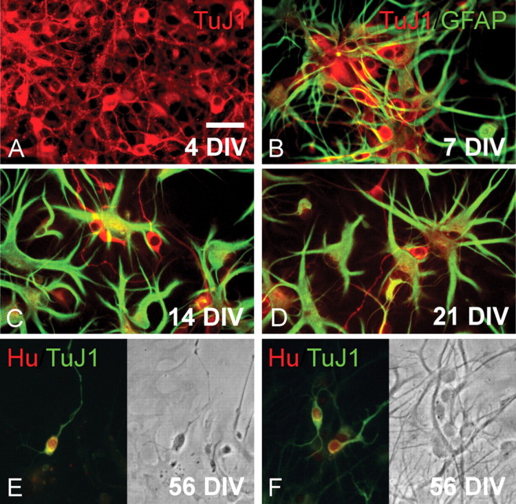
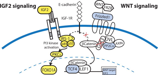
Central neurocytoma (CN) is a rare periventricular tumor, whose derivation, lineage potential, and molecular regulation have been mostly unexplored. We noted that CN cells exhibited an antigenic profile typical of neuronal progenitor cells in vivo, yet in vitro generated neurospheres, divided in response to bFGF (basic fibroblast growth factor), activated the neuroepithelial enhancer of the nestin gene, and gave rise to both neuron-like cells and astrocytes. When CN gene expression was compared with that of both normal adult VZ (ventricular zone) and E/nestin:GFP (green fluorescent protein)-sorted native neuronal progenitors, significant overlap was noted. Marker analysis suggested that the gene expression pattern of CN was that of a proneuronal population; glial markers were conspicuously absent, suggesting that the emergence of astroglia from CN occurred only with passage. The expression pattern of CN was distinguished from that of native progenitor cells by a cohort of differentially expressed genes potentially involved in both the oncogenesis and phenotypic restriction of neurocytoma. These included both IGF2 and several components of its signaling pathway, whose sharp overexpression implicated dysregulated autocrine IGF2 signaling in CN oncogenesis. Both receptors and effectors of canonical wnt signaling, as well as GDF8 (growth differentiation factor 8), PDGF-D, and neuregulin, were differentially overexpressed by CN, suggesting that CN is characterized by the concurrent overactivation of these pathways, which may serve to drive neurocytoma expansion while restricting tumor progenitor phenotype. This strategy of comparing the gene expression of tumor cells to that of the purified native progenitors from which they derive may provide a focused approach to identifying transcripts important to stem and progenitor cell oncogenesis.


Figures

Figure 1.
Figure 1.
Neurocytoma includes mitotic neuroblasts and a minority of postmitotic neuron-like cells. A, Normal adult human ventricular wall. B, A normal ventricular zone, immunostained for βIII-tubulin/TuJ1 (red), GFAP (green), and DAPI (blue). The normal VZ exhibits a well defined layer of GFAP+ subependymal astrocytes. In contrast to neurocytoma, no TuJ1+ cell bodies were observed adjacent to the ventricular wall in normal tissues, although they were observed in adjacent regions of the same sections (inset shows TuJ1+ hippocampal neurons of this section). C, Histological appearance of neurocytoma (hematoxylin and eosin) (region shown in high-power image indicated by asterisk in inset). D–F, Sections of a central neurocytoma, taken from a 20-year-old male patient with a striatal ventricular wall tumor, immunostained for neuronal βIII-tubulin protein using mAb TuJ1 (green). D, E, GFAP+ astrocytes (red) are scattered among the TuJ1+ cells, which comprise the bulk of the tumor mass (green). GFAP+ cells constituted <5% of the total cell population in each of seven tumors examined. Of note, neurocytoma cells were never noted to coexpress both GFAP and TuJ1. F, The majority of the neurocytoma cells expressed musashi protein, a marker of uncommitted neural progenitor cells (green). Scattered among these were less frequent cells expressing Hu protein (red), a marker of terminally committed and postmitotic neurons. The persistence of Hu+ cells within the musashi+ tumor mass likely represents the generation of postmitotic Hu+ neuronal daughter cells from neoplastic musashi+ progenitor cells. Scale bar, 60 μm.
Figure 2.
Figure 2.
Neurocytoma cells can generate both astrocytes and neuron-like cells in culture. A, βIII-tubulin/TuJ1+ (red) cells of neuronal phenotype, in an unpassaged sample of cultured neurocytoma, after 5 d in vitro. B–D, GFAP+ astroglia (green) and TuJ1+ neuroblasts (red), in a second passage culture after 7, 14, and 21 DIV, respectively. Progressive time in vitro was accompanied by a relative expansion of astrocytic derivatives. No cells coexpressing GFAP and TuJ1 were ever seen. E, F, A culture double labeled at 56 DIV for TuJ1/βIII-tubulin and Hu, a postmitotic neuronal protein (Barami et al., 1995), revealed that many of the TuJ1+ cells (green) coexpressed Hu (red nuclei), indicating that neurocytoma cells continued to generate neuron-like cells for extended periods of time in vitro. Scale bar, 30 μm.
Figure 3.
Figure 3.
Neurocytoma exhibits selective glial expansion in vitro. This neurocytoma was resected from a 20-year-old man, in which it presented as an intraventricular mass arising from the striatal wall. A–C, Neuron-like cells in a monolayer culture of this tumor, after 10 DIV. D–F, Subsequent passage revealed the selective expansion of a glial/progenitor phenotype. The generation of occasional neurons (E, arrow) in these cultures suggested their persistent competence as progenitor cells (21 DIV), although their bizarre morphology (D–F) suggested their anaplastic transformation. Scale bar, 25 μm.
Figure 4.
Figure 4.
Neurocytoma can generate passageable neurospheres that give rise to neuron-like cells and astrocytes. A, B, Primary (A) and secondary (B) neurospheres derived from cultured neurocytoma, after monthly passages. C, Neurospheres expressed musashi (green) and nestin (red). D, E, Serial magnifications of secondary neurospheres generated from single CN cells, plated onto collagen to allow the migration of differentiated daughters (2 months in vitro). These secondary neurospheres exhibited emigration of both βIII-tubulin+/TuJ1+ neuron-like cells (green) and GFAP+ astrocytes (red). F, G, AdE/nestin:GFP infection revealed a cohort of GFP+ CN cells that transcriptionally activated the neuroepithelial enhancer of the nestin gene. Scale bars: A, B, 60 μm; C, E, 40 μm; D, 75 μm; F, G, 25 μm.
Figure 5.
Figure 5.
Neurocytoma manifests a loss of imprinting of IGF2 concomitant with IGF2 overexpression. Neurocytoma, immunolabeled for the neuronal marker synaptophysin (red; A), coexpressed IGF-2 (green; B). Virtually all synaptophysin+ neurocytoma cells coexpressed IGF-2. DAPI, 4′,6′-Diamidino-2-phenylindole. C, Southern blotting of neurocytoma genomic DNA coupled with PCR of RNA derived from the same tumor revealed a loss of IGF2 imprinting in the one informative heterozygous case (AB; left). PCR revealed IGF2 transcripts generated from each of the two alleles (right); such a loss of IGF2 imprinting is associated with IGF2 overexpression (Ohlsson, 2004). Scale bar, 40 μm.
Figure 6.
Figure 6.
Dysregulated IGF2 signaling in neurocytoma may regulate tumorigenesis. Differentially overexpressed genes in the IGF2 and wnt pathways, noted in yellow and blue, respectively. Our transcriptional analysis suggests that the genesis of CN from native progenitor cells may involve IGF2 overexpression, possibly via loss of imprinting, and excessive IGF2 receptor activation. We hypothesize that the IGF2 signal may cooperate with frizzled3 signaling to potentiate β-catenin translocation and signaling through TCF/LEF.

References

    1. Akimoto J, Itoh H, Itoh Y, Miwa T. Histogenesis of central neurocytoma: an immunohistochemical and electronmicroscopic study of four cases. No Shinkei Geka. 1995;23:1083–1091. - PubMed
    1. Arsenijevic Y, Villemure JG, Brunet JF, Bloch JJ, Deglon N, Kostic C, Zurn A, Aebischer P. Isolation of multipotent neural precursors residing in the cortex of the adult human brain. Exp Neurol. 2001;170:48–62. - PubMed
    1. Bansal R, Lakhina V, Remedios R, Tole S. Expression of FGF receptors 1, 2, 3 in the embryonic and postnatal mouse brain compared with Pdgfralpha, Olig2 and Plp/dm20: implications for oligodendrocyte development. Dev Neurosci. 2003;25:83–95. - PubMed
    1. Barami K, Iversen K, Furneaux H, Goldman SA. Hu protein as an early marker of neuronal phenotypic differentiation by subependymal zone cells of the adult songbird forebrain. J Neurobiol. 1995;28:82–101. - PubMed
    1. Begley CG, Lipkowitz S, Gobel V, Mahon KA, Bertness V, Green AR, Gough NM, Kirsch IR. Molecular characterization of NSCL, a gene encoding a helix-loop-helix protein expressed in the developing nervous system. Proc Natl Acad Sci USA. 1992;89:38–42. - PMC - PubMed

Publication types