. 2007 Feb;14(2):150-6.
doi: 10.1128/CVI.00285-06. Epub 2006 Nov 29.

Expression of antibodies directed to Paracoccidioides brasiliensis glycosphingolipids during the course of paracoccidioidomycosis treatment

Affiliations

Expression of antibodies directed to Paracoccidioides brasiliensis glycosphingolipids during the course of paracoccidioidomycosis treatment

Silvio Bertini et al. Clin Vaccine Immunol. 2007 Feb.

Abstract

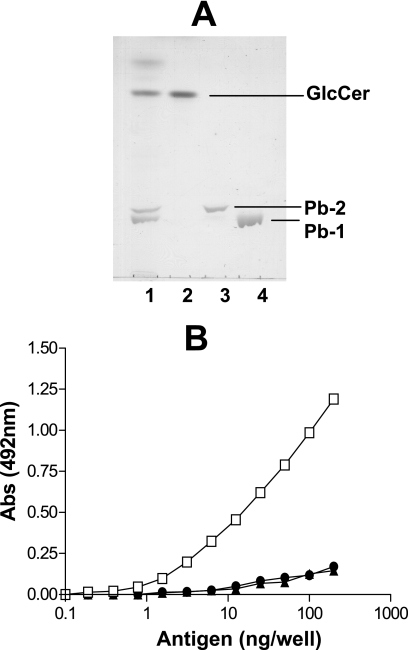
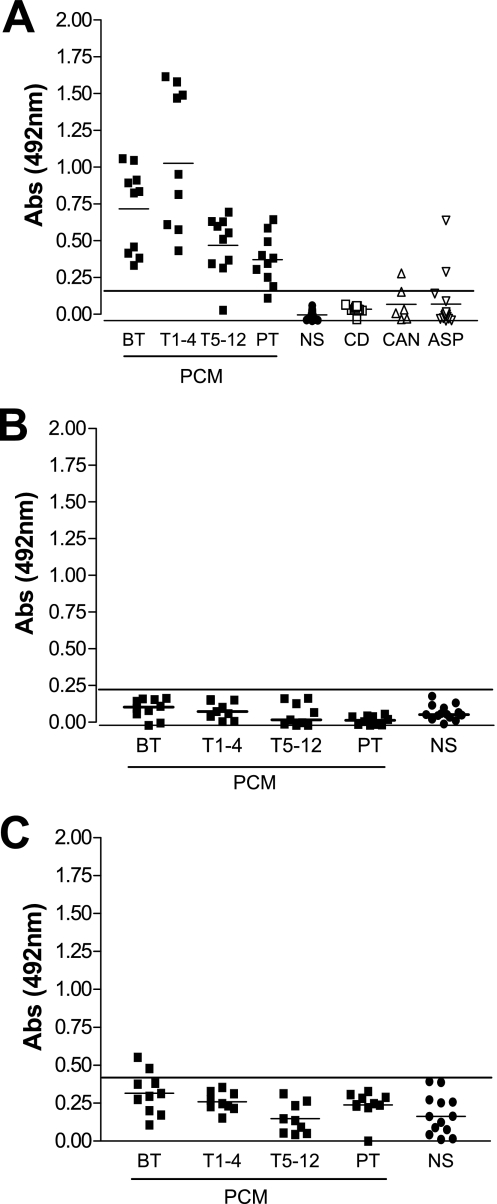
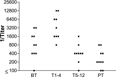
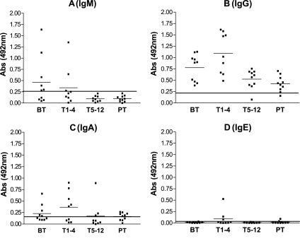
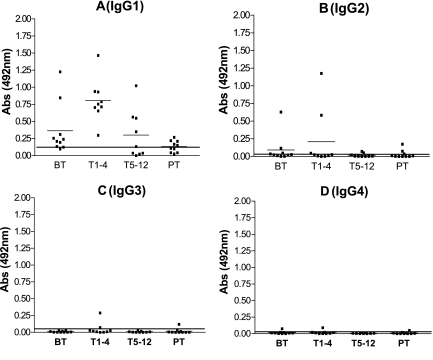
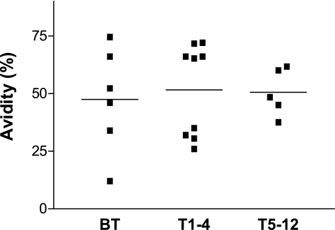
Paracoccidioidomycosis (PCM) is a granulomatous disease caused by the dimorphic fungus Paracoccidioides brasiliensis. The immunoglobulin classes and isotypes of antibodies directed to acidic glycosphingolipids (GSLs) and glucosylceramide of P. brasiliensis were determined by enzyme-linked immunosorbent assay of sera from 31 PCM patients. The reactivities of 38 serum samples were analyzed by considering the stage of treatment: before antifungal treatment (n = 10), during 1 to 4 months of treatment (T1-4; n = 9), during 5 to 12 months of treatment (T5-12; n = 9), and posttreatment (PT; n = 10). Sera from healthy subjects (n = 12) were used as controls. Only the GSL Pb-1 antigen, which presents the carbohydrate structure Galfbeta1-6(Manalpha1-3)Manbeta1, was reactive with the PCM patient sera. The PCM patient sera did not react with Pb-2, which lacks the Galf residue and which is considered the biosynthetic precursor of Pb-1, indicating that the Galf residue is essential for antibody reactivity. The Pb-1 glycolipid from nontreated patients elicited a primary immune response with immunoglobulin M (IgM) production and subsequent switching to IgG1 production. The IgG1 titer increased after the start of antifungal treatment (T1-4 group), and general decreases in the anti-Pb-1 antibody titers were observed after 5 months of treatment (T5-12 and PT groups). The Pb-1 antigen, an acidic GSL with terminal Galf residue, has potential application as an elicitor of the host immune response in patients with PCM.


Figures

FIG. 1.
FIG. 1.
Purified GSLs from P. brasiliensis and their immunogenicities. (A) HPTLC pattern of purified GSLs from P. brasiliensis obtained by using the solvent chloroform-methanol-CaCl2 (0.02%) and staining with orcinol-H2SO4. Lane 1, total P. brasiliensis GSLs; lane 2, purified GlcCer; lane 3, purified Pb-2; lane 4, purified Pb-1. (B) Reactivities of sera from PCM patients (n = 5; diluted 100 times) with Pb-1 (squares), Pb-2 (triangles), and GlcCer (circles) purified from P. brasiliensis by ELISA before treatment. Various concentrations of GSLs were adsorbed on 96-well plates, and the plates were blocked and incubated overnight at 4°C with pooled patient sera (n = 5). The amount of Ig bound to each GSL was determined by using biotinylated anti-IgG (heavy and light chains), as described in Materials and Methods. Abs, absorbance.
FIG. 2.
FIG. 2.
Reactivities of sera of patients with PCM and Chagas' disease with various GSLs. Samples (∼100 ng) of Pb-1 (A), Pb-2 (B), and GlcCer (C) were adsorbed on 96-well plates and incubated with sera from PCM patients (diluted 100 times) taken at various stages of treatment (BT, n = 10; T1-4, n = 9; T5-12, n = 9; PT, n = 10) or with sera from patients with Chagas' disease (CD; n = 9), patients with invasive candidiasis (CAN; n = 6), or patients with pulmonary aspergillosis (ASP; n = 14). The amount of Ig bound to each GSL was determined by using biotinylated anti-IgG (heavy and light chains), as described in Materials and Methods. Serum reactivity was analyzed by ELISA. Short horizontal lines, mean absorbance for each group; long horizontal lines, cutoff determined for sera from healthy subjects (NS; n = 12). Abs, absorbance.
FIG. 3.
FIG. 3.
Pb-1 antibody titers in PCM patient serum. Purified Pb-1 antigen (100 ng/well) was adsorbed on a 96-well plate and incubated with serially diluted sera from the PCM patient groups, as defined in the legend to Fig. 2. The amount of bound antibody was determined by ELISA with biotinylated mouse anti-human IgG (heavy and light chains). The titer was determined as the last serum dilution with an absorbance value higher than 0.1.
FIG. 4.
FIG. 4.
Expression of Ig directed to Pb-1 antigen in PCM patient sera. Purified Pb-1 (100 ng/well) was adsorbed on a 96-well plate and incubated with sera from the PCM patient groups, as defined in the legend to Fig. 2. The amount of bound antibody was determined by ELISA with biotinylated mouse anti-human Ig, as described in Materials and Methods. (A) IgM (μ chain); (B) IgG (γ chain); (C) IgA; (D) IgE. The short and the long horizontal lines are as defined in the legend to Fig. 2. Abs, absorbance.
FIG. 5.
FIG. 5.
Expression of IgG subclass directed to Pb-1 antigen in PCM patient sera. Purified Pb-1 (100 ng/well) was adsorbed on a 96-well plate and incubated with sera from the PCM patient groups, as defined in the legend to Fig. 2. The amount of bound antibody was determined by ELISA with biotinylated mouse anti-human Ig, as described in Materials and Methods. (A) IgG1; (B) IgG2; (C) IgG3; (D) IgG4. The short and the long horizontal lines are as defined in the legend to Fig. 2. Abs, absorbance.
FIG. 6.
FIG. 6.
Avidity of anti-Pb-1 IgG antibody present in PCM patient sera. Sera from patients with chronic PCM were serially diluted and incubated in 96-well plates preadsorbed with 100 ng Pb-1. The avidities of the anti-Pb-1 antibodies, defined as the capacity to remain bound to the Pb-1 antigen in the presence of 6.0 M urea in PBS, were determined by ELISA. Antibody binding was determined with biotinylated anti-human IgG (γ chain) as the secondary antibody, as described in Materials and Methods. Bars, mean absorbance value for each group.

References

    1. Baida, H., P. J. C. Biselli, M. Juvenale, G. J. S. Del Negro, M. J. S. Mendes-Giannini, A. J. S. Duarte, and G. Benard. 1999. Differential antibody isotype expression to the major Paracoccidioides brasiliensis antigen in juvenile and adult form paracoccidioidomycosis. Microbes Infect. 1:273-278. - PubMed
    1. Biselli, P. J. C., M. Juvenale, M. J. S. Mendes-Giannini, A. J. S. Durte, and G. Benard. 2001. IgE antibody response to the main component of Paracoccidioides brasiliensis in patient with paracoccidioidomycosis. Med. Mycol. 39:475-478. - PubMed
    1. Brummer, E., E. Castaneda, and A. Restrepo. 1993. Paracoccidioidomycosis: an update. Clin. Microbiol. Rev. 6:89-117. - PMC - PubMed
    1. De Camargo, Z. P., and M. F. Franco. 2000. Current knowledge on pathogenesis and immunodiagnosis of paracoccidioidomycosis. Rev. Iberoam. Micol. 17:41-48. - PubMed
    1. De Camargo, Z. P., C. Unterkircher, S. P. Campoy, and L. R. Travassos. 1988. Production of Paracoccidioides brasiliensis exoantigens for immunodiffusion test. J. Clin. Microbiol. 26:2147-2151. - PMC - PubMed

Publication types