

Inhibition of inducible Nitric Oxide Synthase by a mustard gas analog in murine macrophages
- PMID: 17137498
- PMCID: PMC1698482
- DOI: 10.1186/1471-2121-7-39
Inhibition of inducible Nitric Oxide Synthase by a mustard gas analog in murine macrophages
Abstract
Background: 2-Chloroethyl ethyl sulphide (CEES) is a sulphur vesicating agent and an analogue of the chemical warfare agent 2,2'-dichlorodiethyl sulphide, or sulphur mustard gas (HD). Both CEES and HD are alkylating agents that influence cellular thiols and are highly toxic. In a previous publication, we reported that lipopolysaccharide (LPS) enhances the cytotoxicity of CEES in murine RAW264.7 macrophages. In the present investigation, we studied the influence of CEES on nitric oxide (NO) production in LPS stimulated RAW264.7 cells since NO signalling affects inflammation, cell death, and wound healing. Murine macrophages stimulated with LPS produce NO almost exclusively via inducible nitric oxide synthase (iNOS) activity. We suggest that the influence of CEES or HD on the cellular production of NO could play an important role in the pathophysiological responses of tissues to these toxicants. In particular, it is known that macrophage generated NO synthesised by iNOS plays a critical role in wound healing.
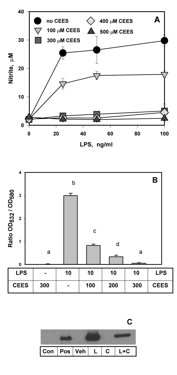
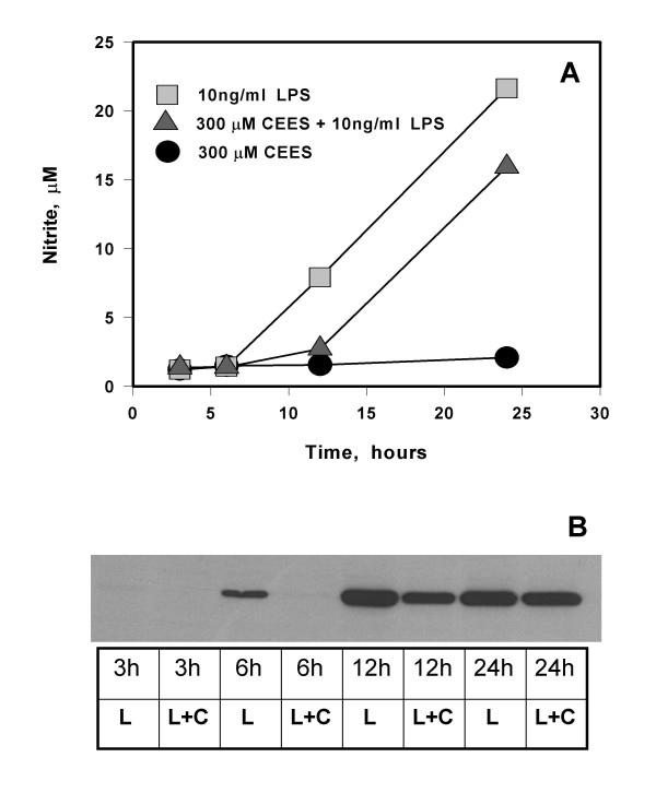
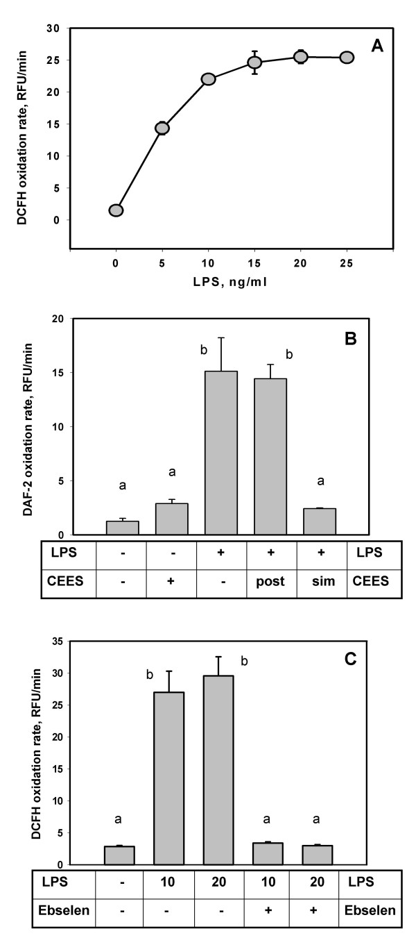
Results: We initially confirmed that in LPS stimulated RAW264.7 macrophages NO is exclusively generated by the iNOS form of nitric oxide synthase. CEES treatment inhibited the synthesis of NO (after 24 hours) in viable LPS-stimulated RAW264.7 macrophages as measured by either nitrite secretion into the culture medium or the intracellular conversion of 4,5-diaminofluorescein diacetate (DAF-2DA) or dichlorofluorescin diacetate (DCFH-DA). Western blots showed that CEES transiently decreased the expression of iNOS protein; however, treatment of active iNOS with CEES in vitro did not inhibit its enzymatic activity
Conclusion: CEES inhibits NO production in LPS stimulated macrophages by decreasing iNOS protein expression. Decreased iNOS expression is likely the result of CEES induced alteration in the nuclear factor kappa B (NF-kappaB) signalling pathway. Since NO can act as an antioxidant, the CEES induced down-regulation of iNOS in LPS-stimulated macrophages could elevate oxidative stress. Since macrophage generated NO is known to play a key role in cutaneous wound healing, it is possible that this work has physiological relevance with respect to the healing of HD induced skin blisters.
Figures
Similar articles
-
The influence of N-acetyl-L-cysteine on oxidative stress and nitric oxide synthesis in stimulated macrophages treated with a mustard gas analogue.BMC Cell Biol. 2008 Jun 20;9:33. doi: 10.1186/1471-2121-9-33. BMC Cell Biol. 2008. PMID: 18570648 Free PMC article.
-
Inhibition of inducible nitric oxide synthase by Acanthopanax senticosus extract in RAW264.7 macrophages.J Ethnopharmacol. 2008 Jul 23;118(2):231-6. doi: 10.1016/j.jep.2008.04.003. Epub 2008 Apr 11. J Ethnopharmacol. 2008. PMID: 18486372
-
[Regulation of lipopolysaccharide-induced inducible nitric oxide synthase gene expression by protein kinase C].Zhonghua Yi Xue Za Zhi. 2002 Nov 10;82(21):1488-92. Zhonghua Yi Xue Za Zhi. 2002. PMID: 12509913 Chinese.
-
Role of inducible nitric oxide synthase in the regulation of leucocyte recruitment.Clin Sci (Lond). 2001 Jan;100(1):1-12. Clin Sci (Lond). 2001. PMID: 11115411 Review.
-
Efficacy of glutathione in ameliorating sulfur mustard analog-induced toxicity in cultured skin epidermal cells and in SKH-1 mouse skin in vivo.J Pharmacol Exp Ther. 2011 Feb;336(2):450-9. doi: 10.1124/jpet.110.173708. Epub 2010 Oct 25. J Pharmacol Exp Ther. 2011. PMID: 20974699 Free PMC article. Review.
Cited by
-
The influence of N-acetyl-L-cysteine on oxidative stress and nitric oxide synthesis in stimulated macrophages treated with a mustard gas analogue.BMC Cell Biol. 2008 Jun 20;9:33. doi: 10.1186/1471-2121-9-33. BMC Cell Biol. 2008. PMID: 18570648 Free PMC article.
-
Sulfur mustard toxicity following dermal exposure: role of oxidative stress, and antioxidant therapy.J Burns Wounds. 2007 Oct 30;7:e7. J Burns Wounds. 2007. PMID: 18091984 Free PMC article.
-
Selective targeting of selenocysteine in thioredoxin reductase by the half mustard 2-chloroethyl ethyl sulfide in lung epithelial cells.Chem Res Toxicol. 2010 Jun 21;23(6):1045-53. doi: 10.1021/tx100040k. Chem Res Toxicol. 2010. PMID: 20345183 Free PMC article.
-
A role for mitochondrial oxidative stress in sulfur mustard analog 2-chloroethyl ethyl sulfide-induced lung cell injury and antioxidant protection.J Pharmacol Exp Ther. 2009 Mar;328(3):732-9. doi: 10.1124/jpet.108.145037. Epub 2008 Dec 8. J Pharmacol Exp Ther. 2009. PMID: 19064720 Free PMC article.
-
Oxidants and antioxidants in sulfur mustard-induced injury.Ann N Y Acad Sci. 2010 Aug;1203:92-100. doi: 10.1111/j.1749-6632.2010.05605.x. Ann N Y Acad Sci. 2010. PMID: 20716289 Free PMC article. Review.
References
-
- Smith KJ, Skelton H. Chemical warfare agents: their past and continuing threat and evolving therapies. Part I of II. Skinmed. 2003;2:215–221. - PubMed
Publication types
MeSH terms
Substances
LinkOut - more resources
Full Text Sources
Miscellaneous
