. 2007 Jan;34(1):48-58.
doi: 10.1016/j.mcn.2006.10.004. Epub 2006 Nov 29.

SR protein 9G8 modulates splicing of tau exon 10 via its proximal downstream intron, a clustering region for frontotemporal dementia mutations

Affiliations

SR protein 9G8 modulates splicing of tau exon 10 via its proximal downstream intron, a clustering region for frontotemporal dementia mutations

Lei Gao et al. Mol Cell Neurosci. 2007 Jan.

Abstract

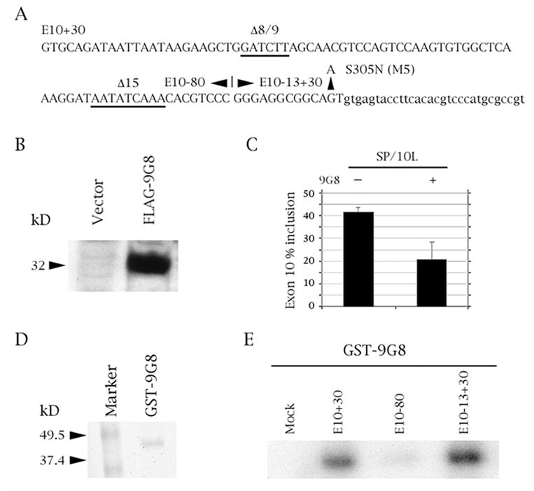
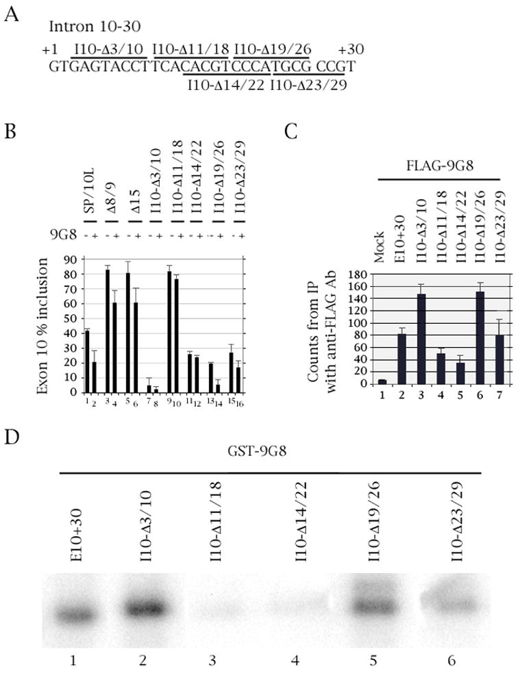
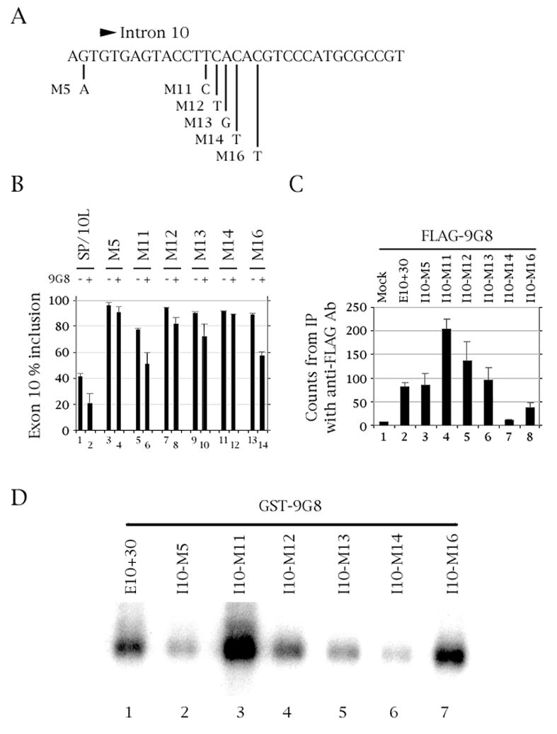
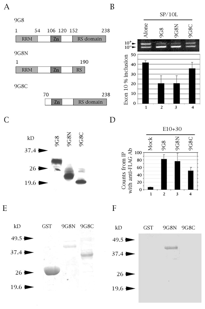
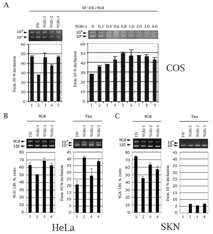
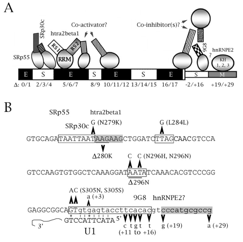
The microtubule-associated protein tau is important to normal neuronal function in the mammalian nervous system. Aggregated tau is the major component of neurofibrillary tangles (NFTs), present in several neurodegenerative diseases, including Alzheimer's and frontotemporal dementia with Parkinsonism (FTDP). Splicing misregulation of adult-specific exon 10 results in expression of abnormal ratios of tau isoforms, leading to FTDP. Positions +3 to +16 of the intron downstream of exon 10 define a clustering region for point mutations that are found in FTDP. The serine/arginine-rich (SR) factor 9G8 strongly inhibits inclusion of tau exon 10. In this study, we established that 9G8 binds directly to this clustering region, requires a wild-type residue at position +14 to inhibit exon inclusion, and RNAi constructs against 9G8 increase exon 10 inclusion. These results indicate that 9G8 plays a key role in regulation of exon 10 splicing and imply a pathogenic role in neurodegenerative diseases.


Figures

Fig. 1
Fig. 1
SR factor 9G8 inhibits splicing of tau exon 10 by binding to the proximal downstream intron. (A) Tau exon 10 (uppercase) and its downstream intron (lowercase). Relevant exonic deletions are underlined and point mutation S305N (M5) is shown. The boundaries of the three riboprobe constructs are indicated. E10+30 contains exon 10 plus 30 nucleotides of its downstream intron, E10-80 contains the 80 5’-most nucleotides of exon 10, E10-13+30 contains the 13 3’most nucleotides of exon 10 plus 30 nucleotides of its downstream intron. (B, D) Expression of FLAG-9G8 and GST-9G8 fusion proteins, respectively detected by (B) Anti-FLAG monoclonal antibody and (D) Coomassie blue staining. (C) RT-PCR of SP/10L in COS cells in the absence and presence of 9G8. The RT-PCR products come from 1:1 co-transfections of SP/10L and FLAG-9G8. The identities of the spliced species are indicated. Primer pair: SPL-LS/SPL-LN. % exon inclusion was calculated by scanning the bands from three independent transfections and measuring their areas using the OneDscan analysis program. (E) UV-crosslinking of GST-9G8 to the three riboprobes indicated in (A). Equal amounts of the fusion protein were present in each experiment.
Fig. 2
Fig. 2
Deletion of residues 11-18 of the intron downstream of tau exon 10 abolishes 9G8 action and binding. (A) Nucleotides +1 to +30 of the intron downstream of tau exon 10. The deletions are demarcated by bars above or below the sequence. (B) RT-PCR of wild-type and deleted SP/10L in COS cells in the absence and presence of 9G8. The RT-PCR products come from 1:1 co-transfections of tau constructs and FLAG-9G8. Exon ratio calculations, primers and graph conventions are as in Fig.1C. (C) 32P-labeled riboprobes containing wild-type or deleted exon 10 were incubated with extracts from COS cells transfected with FLAG-9G8 and were immunoprecipitated by anti-FLAG monoclonal antibody. Amounts of riboprobe bound to 9G8 protein were calculated relative to the nonspecific binding of the mock vector transfection (means ± SD of three analyses). (D) UV-crosslinking of GST-9G8 to the deletion substrates indicated in (A). Equal amounts of the fusion protein were present in each experiment.
Fig. 3
Fig. 3
Mutation of residue +14 of the intron downstream of tau exon 10 abolishes 9G8 action and binding. (A) The last three nucleotides of exon 10 and nucleotides +1 to +30 of the intron downstream of tau exon 10. The point mutations are indicated. (B) RT-PCR of wild-type and point-mutated SP/10L in COS cells in the absence and presence of 9G8. The RT-PCR products come from 1:1 co-transfections of tau constructs and FLAG-9G8. Exon ratio calculations, primers and graph conventions are as in Fig. 1C. (C) 32P-labeled riboprobes containing wild-type or point-mutated exon 10 were incubated with extracts from COS cells transfected with FLAG-9G8 and were immunoprecipitated by anti-FLAG monoclonal antibody. Amounts of riboprobe bound to 9G8 protein were calculated relative to the nonspecific binding of the mock vector transfection (means ± SD of three analyses). (D) UV-crosslinking of GST-9G8 to the deletion substrates indicated in (A). Equal amounts of the fusion protein were present in each experiment.
Fig. 4
Fig. 4
The RRM domain of 9G8 is required for inhibition of tau exon 10 splicing and binds to tau exon 10. (A) Diagram of 9G8 deletion variants. The amino acid number, RNA recognition motif (RRM), zinc knuckle (Zn) and serine/arginine-rich region (RS) are indicated. (B) RT-PCR of SP/10L in COS cells in the presence of full-length and deletion variants of 9G8. The RT-PCR products come from 1:1 co-transfections of SP/10L and FLAG-9G8 fusion constructs. Exon ratio calculations, primers and graph conventions are as in Fig. 1C. (C, E) Expression of FLAG-9G8 and GST-9G8 deleted fusion proteins, respectively detected by (C) Anti-FLAG monoclonal antibody and (E) Coomassie blue staining. (D) 32P-labeled riboprobes containing wild-type exon 10 were incubated with extracts from COS cells transfected with FLAG-9G8 variants and were immunoprecipitated by anti-FLAG monoclonal antibody. Amounts of riboprobe bound to 9G8 protein were calculated relative to the nonspecific binding of the mock vector transfection (means ± SD of three analyses). (F) UV-crosslinking of E10+30 to the 9G8 variants indicated in (A). Equal amounts of the fusion protein were present in each experiment.
Fig. 5
Fig. 5
Three RNAi constructs against 9G8 decrease expression of endogenous 9G8 and counteract inhibition of endogenous tau exon 10 splicing in a sequence- and dosage-dependent manner. (A) Establishment of RNAi construct efficacy. RT-PCR of SP/10L and three 9G8 RNAi constructs in COS cells. Primer pair: SPL-LS/SPL-LN. (Left panel) 1 ug of SP/10L and 1 ug of FLAG-9G8 were co-transfected into the cells together with 1 ug of the RNAi constructs (9G8i-1, 2, 3) or the RNAi vector pFIV H1/U6 (FIV). (Right panel) RT-PCR of SP/10L with increasing amounts of construct 9G8i-1. 1 ug of SP/10L and 1ug of FLAG-9G8 were co-transfected in COS cells with the indicated amounts of RNAi-1 plasmid (in ug). (B, C) Effects of the three 9G8 RNAi constructs on endogenous 9G9 and tau in (B) HeLa and (C) SKN cells. (B, C, Left panels) Quantitative RT-PCR of endogenous 9G8. The % ratio of 9G8 to 18S is indicated. Primer pair: 9G8ex3S/9G8ex8N. (B, C, Right panels) Quantitative RT-PCR of tau exon 10. The % of tau exon 10 inclusion is indicated. Primer pair: HT7S3/HT11N. (A-C) Exon ratio calculations and graph conventions are as in Fig. 1C.
Fig. 6
Fig. 6
Speculative model of splicing regulation for two elements of tau exon 10. (A) Schematic representation of splicing factor interactions with exon 10. The exon is denoted by thick boxes, its downstream intron by thin boxes. The deletion nomenclature is according to D’Souza and Schellenberg (2000), Wang et al. (2005). The behavior of each region as delimited by the deletions is indicated by E (enhancer), S (silencer) or M (modifier). For the factors, circles represent RRM domains, squares represent RS domains. The model of the exonic silencer and enhancer at the 5’ end of the exon comes from Wang et al. (2005), with additional information from D’Souza and Schellenberg (2000), Jiang et al. (2003), Kondo et al. (2004). The model of the elements and factors in the proximal downstream intron come from this study and from Broderick et al. (2004). Question marks imply an unknown or hypothesized interaction. 5’ end: SRp30c and SRp55 bind to region E10-Δ2/3/4, whereas htra2beta1 binds to region E10-Δ5/6. The two inhibitors interact (or sterically interfere) with the RS1 domain of htra2beta1; its RS2 domain may interact with a putative co-activator which may bind to either E10-Δ6/7 or E10-Δ10/11/12. 3’ end: 9G8 binds to residues +11 to +18 but its influence is also abolished in mutant M5. 9G8 is probably part of a larger complex whose action may be modified or antagonized by hnRNPE2, which moderately activates splicing of exon 10. (B) Context of FTDP mutations that affect splicing within the cis regulatory elements depicted in (A). The sequence of exon 10 and its proximal downstream intron is shown. Exonic sequences are in uppercase, intronic in lowercase. FTDP point mutations and deletions are indicated. The boxed regions define enhancers (gray) or silencers (white). Also shown are the factors known or suspected to interact with regulatory cis elements, and the complementarity of the 5’ splice site with the U1 snRNA. Lines are regular Crick/Watson pairs, dots are G-T pairs.

Similar articles

Cited by

References

    1. Andreadis A. Tau gene alternative splicing: expression patterns, regulation and modulation of function in normal brain and neurodegenerative diseases. Biochem Biophys Acta. 2005;1739:91–103. - PubMed
    1. Brandt R, Léger J, Lee G. Interaction of tau with the neural plasma membrane mediated by tau's amino-terminal projection domain. J Cell Biol. 1995;131:1327–1340. - PMC - PubMed
    1. Brandt R, Hundelt M, Shahani N. Tau alteration and neuronal degeneration in tauopathies: mechanisms and models. Biochem Biophys Acta. 2005;1739:331–354. - PubMed
    1. Broderick JA, Wang J, Andreadis A. Heterogeneous nuclear ribonucleoprotein E2 binds to tau exon 10 and moderately activates its splicing. Gene. 2004;331:107–114. - PubMed
    1. Cáceres JF, Misteli T, Screaton GR, Spector DL, Krainer AR. Role of the modular domains of SR proteins in subnuclear localization and alternative splicing specificity. J Cell Biol. 1997;138:225–238. - PMC - PubMed

Publication types

MeSH terms