. 2006 Dec 12;103(50):19188-93.
doi: 10.1073/pnas.0607466103. Epub 2006 Nov 30.

GABA and Gi/o differentially control circadian rhythms and synchrony in clock neurons

Affiliations

GABA and Gi/o differentially control circadian rhythms and synchrony in clock neurons

Sara J Aton et al. Proc Natl Acad Sci U S A. .

Abstract

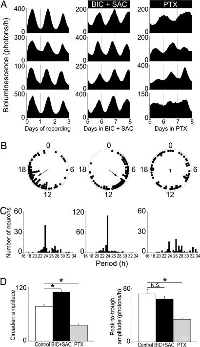
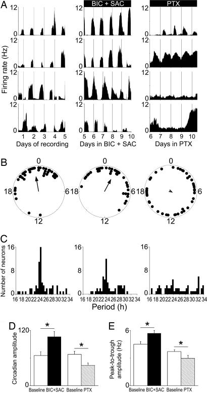
Neurons in the mammalian suprachiasmatic nuclei (SCN) generate daily rhythms in physiology and behavior, but it is unclear how they maintain and synchronize these rhythms in vivo. We hypothesized that parallel signaling pathways in the SCN are required to synchronize rhythms in these neurons for coherent output. We recorded firing and clock-gene expression patterns while blocking candidate signaling pathways for at least 8 days. GABA(A) and GABA(B) antagonism increased circadian peak firing rates and rhythm precision of cultured SCN neurons, but G(i/o) did not impair synchrony or rhythmicity. In contrast, inhibiting G(i/o) with pertussis toxin abolished rhythms in most neurons and desynchronized the population, phenocopying the loss of vasoactive intestinal polypeptide (VIP). Daily VIP receptor agonist treatment restored synchrony and rhythmicity to VIP(-/-) SCN cultures during continuous GABA receptor antagonism but not during G(i/o) blockade. Pertussis toxin did not affect circadian cycling of the liver, suggesting that G(i/o) plays a specialized role in maintaining SCN rhythmicity. We conclude that endogenous GABA controls the amplitude of SCN neuronal rhythms by reducing daytime firing, whereas G(i/o) signaling suppresses nighttime firing, and it is necessary for synchrony among SCN neurons. We propose that G(i/o), not GABA activity, converges with VIP signaling to maintain and coordinate rhythms among SCN neurons.


Conflict of interest statement

The authors declare no conflict of interest.

Figures

Fig. 1.
Fig. 1.
TTX and PTX, not GABA receptor antagonism, damp circadian rhythms from rat Per1::luc SCN slices. (A) Representative traces of detrended bioluminescence from individual SCN cultures in each treatment group. Bars indicate the duration of treatment. (B) Mean peak-to-trough amplitudes (± SEM) of rhythms show that TTX- and PTX-treated slices damped significantly compared with untreated and BIC+SAC-treated slices (n = at least 6 explants per point). Data were normalized to the amplitude of the last cycle before drug treatment. Lines show best-fit result of three models described in Results.
Fig. 2.
Fig. 2.
PTX, not GABA receptor antagonism, disrupts rhythms and synchrony of PER2::LUC rhythms in SCN neurons. (A) Representative PER2::LUC traces from individual neurons within mouse SCN slices under control conditions (Left), on days 5–8 of treatment with BIC+SAC (Center), and on days 5–8 of treatment with PTX (Right). (B) Representative Rayleigh plots of all rhythmic neurons within a SCN slice from each treatment group. The bioluminescence acrophase of each neuron (filled circles) and the mean phase of all neurons (arrow) are plotted for the last 24 h of each treatment. The arrow length is proportional to the magnitude of the phase clustering (r), ranging from 0 (randomly phased) to 1 (peaking at the same time). Whereas control and BIC+SAC-treated neurons maintained phase synchrony (n = 89, r = 0.63, and n = 85, r = 0.56, respectively; P < 0.001, Rayleigh test), PTX-treated neurons peaked randomly (n = 73, r = 0.16; P > 0.1). (C) Period distributions for all neurons recorded in each treatment group. PTX significantly broadened the period distribution of neurons compared with control and BIC+SAC-treated neurons (P < 0.00005, Brown–Forsythe and Levene test). (D) BIC+SAC increased, and PTX decreased the daily precision (as measured by circadian amplitude) of PER2::LUC bioluminescence rhythms in individual SCN neurons relative to controls (*, P < 0.05, ANOVA with Scheffé post hoc test). (E) Compared with controls, PTX decreased (P < 0.05), and BIC+SAC (P > 0.05) did not affect, the peak-to-trough amplitude of bioluminescence rhythms.
Fig. 3.
Fig. 3.
PTX, not GABA receptor antagonism, impairs firing rhythmicity and synchrony between neurons. (A) Representative firing-rate rhythms for individual SCN neurons within a control culture (Left), over the last 5 days of a 10-day BIC+SAC treatment (Center), and on the last 5 days of a 10-day PTX treatment (Right). Neurons reliably peaked at similar times during control and BIC+SAC treatment, but they were less likely to maintain rhythms, they showed lower-amplitude rhythms, and they had unstable phase relationships during PTX treatment. (B) Peak phases of all rhythmic neurons within representative SCN cultures on the last day of baseline recording (Left), on the 10th day of BIC+SAC treatment (Center), and on the 10th day of PTX treatment (Right). Rayleigh distributions of peak phases before treatment (n = 45, r = 0.66) and during BIC+SAC treatment (n = 37, r = 0.59) were statistically nonrandom (P < 0.001), but they were random during PTX treatment (n = 41, r = 0.09; P > 0.6). (C) Period distributions for all rhythmic neurons were similar before (Left) and during (Center) BIC+SAC treatment (P > 0.4), and they were significantly broadened by PTX (Right; P < 0.00001). (D) Circadian amplitudes were greater during BIC+SAC treatment than during baseline recording (*, P < 0.05) and reduced during PTX treatment relative to baseline (P < 0.05). (E) As a result of increased daily peak firing rate (P < 0.05), the peak-to-trough amplitude increased significantly during BIC+SAC treatment (P < 0.05). As a result of increased daily minimum firing rate (P < 0.00001), the peak-to-trough amplitude decreased significantly during PTX treatment (Right; P < 0.05).

References

    1. Hastings MH, Herzog ED. J Biol Rhythms. 2004;19:400–413. - PubMed
    1. Aton SJ, Herzog ED. Neuron. 2005;48:531–534. - PMC - PubMed
    1. Yamaguchi S, Isejima H, Matsuo T, Okura R, Yagita K, Kobayashi M, Okamura H. Science. 2003;302:1408–1412. - PubMed
    1. Honma S, Shirakawa T, Nakamura W, Honma K-I. Neurosci Lett. 2000;294:113–116. - PubMed
    1. Aton SJ, Colwell CS, Harmar AJ, Waschek J, Herzog ED. Nat Neurosci. 2005;8:476–483. - PMC - PubMed

Publication types

LinkOut - more resources