. 2006 Nov;18(11):3332-45.
doi: 10.1105/tpc.106.043588. Epub 2006 Nov 30.

A ferroxidation/permeation iron uptake system is required for virulence in Ustilago maydis

Affiliations

A ferroxidation/permeation iron uptake system is required for virulence in Ustilago maydis

Heiko Eichhorn et al. Plant Cell. 2006 Nov.

Abstract

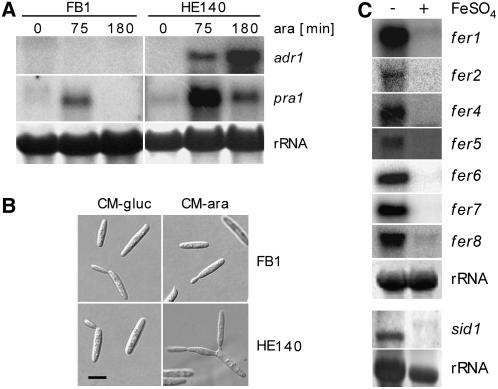
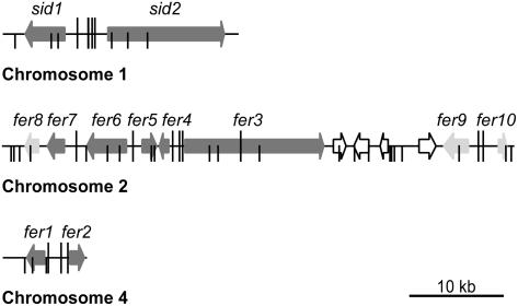
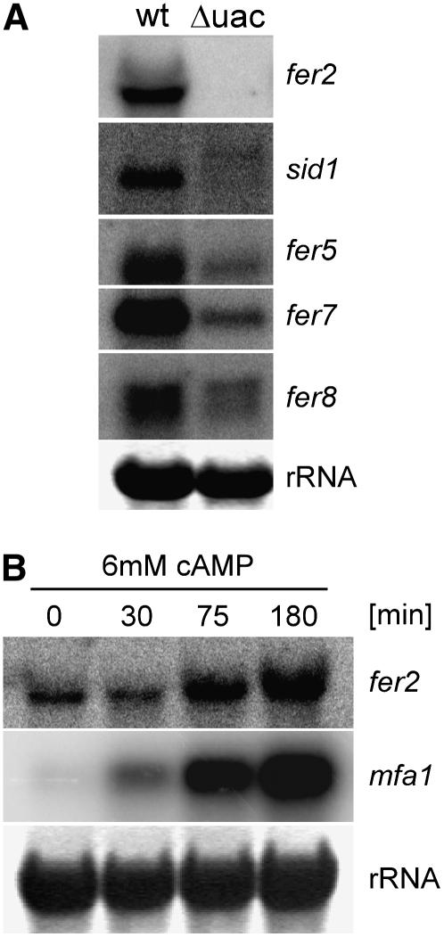
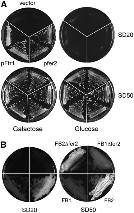
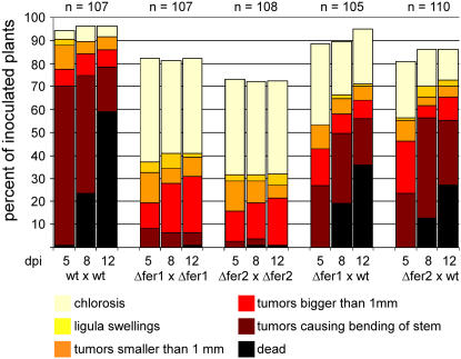
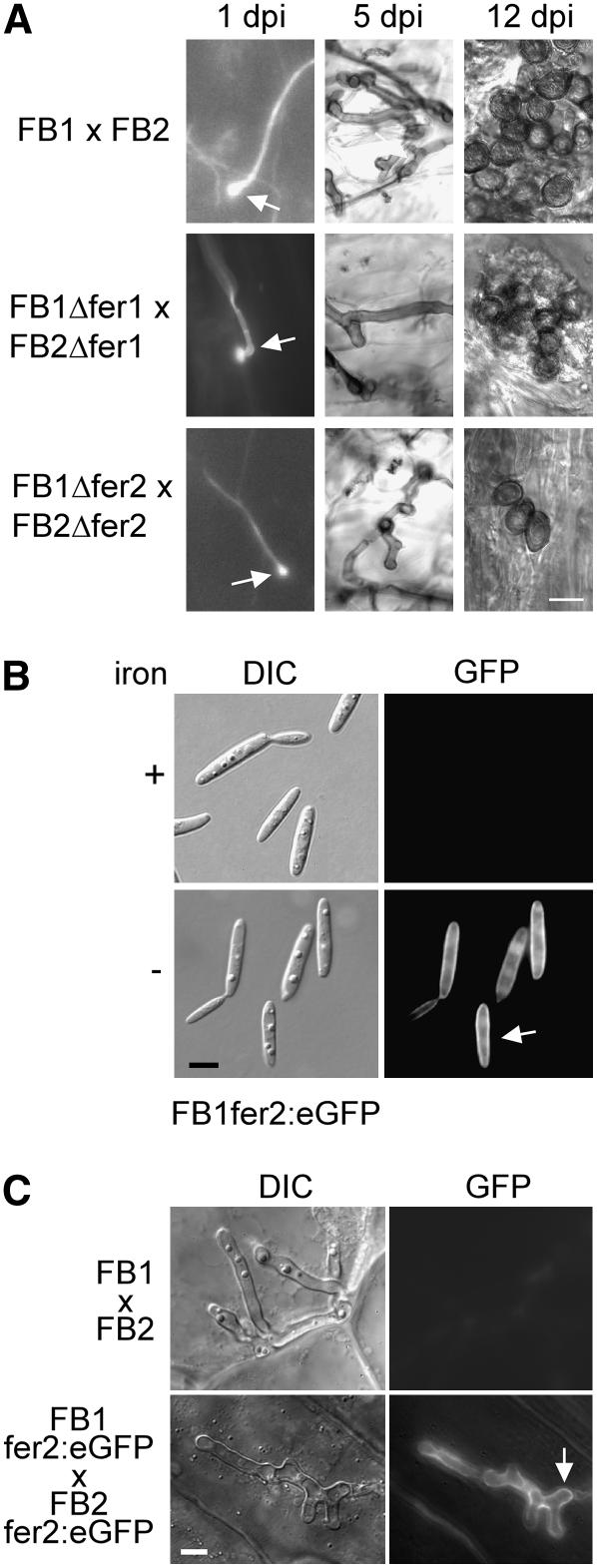
In the smut fungus Ustilago maydis, a tightly regulated cAMP signaling cascade is necessary for pathogenic development. Transcriptome analysis using whole genome microarrays set up to identify putative target genes of the protein kinase A catalytic subunit Adr1 revealed nine genes with putative functions in two high-affinity iron uptake systems. These genes locate to three gene clusters on different chromosomes and include the previously identified complementing siderophore auxotroph genes sid1 and sid2 involved in siderophore biosynthesis. Transcription of all nine genes plus three additional genes associated with the gene clusters was also coregulated by iron through the Urbs1 transcription factor. Two components of a high-affinity iron uptake system were characterized in more detail: fer2, encoding a high-affinity iron permease; and fer1, encoding an iron multicopper oxidase. Fer2 localized to the plasma membrane and complemented an ftr1 mutant of Saccharomyces cerevisiae lacking a high-affinity iron permease. During pathogenic development, fer2 expression was confined to the phase of hyphal proliferation inside the plant. fer2 as well as fer1 deletion mutants were strongly affected in virulence. These data highlight the importance of the high-affinity iron uptake system via an iron permease and a multicopper oxidase for biotrophic development in the U. maydis/maize (Zea mays) pathosystem.


Figures

Figure 1.
Figure 1.
Characterization of Strain HE140 and Repression of Iron Uptake Cluster Genes by Iron. (A) Induction of the crg1 promoter in U. maydis strain HE140 (a1b1[Pcrg1:adr1]ips) by arabinose (ara) induces adr1 and pra1 gene expression. Total RNA (10 μg) from the wild type (FB1) and strain HE140 isolated at 0, 75, and 180 min after the shift to arabinose-containing medium was loaded in each lane and probed successively with pra1 and adr1. In the top panel, the same filter was probed successively with the genes indicated. In the middle panel, a separate filter was probed with sid1. Methylene blue staining of the rRNA is shown in the bottom panel as a loading control for the two separate filters used. (B) Arabinose induction of U. maydis strain HE140 leads to unipolar budding. Light microscopy is shown for wild-type FB1 and HE140 strains grown in complete medium (CM) (Holliday, 1974) supplemented with 1% glucose (left) and shifted for 180 min to CM containing 1% arabinose (right). Bar = 5 μm. (C) Iron represses the expression of genes localized in the high-affinity iron uptake clusters. Total RNA (10 μg) from wild-type strain FB1 grown in minimal medium (Sundström, 1964) without (−) and with (+) the addition of 10 μM FeSO4 was used in each lane. Blots were successively probed with gene-specific probes as indicated at right. Methylene blue staining of the rRNA is shown as a loading control at bottom.
Figure 2.
Figure 2.
Physical Map of Three Coregulated Iron Uptake Clusters. The coregulated genes localize to clusters on three chromosomes of U. maydis. Coding regions are represented by arrows indicating the direction of transcription. Dark gray arrows, coregulated genes involved in iron uptake identified by arabinose induction of HE140; light gray arrows, additional genes identified as being coregulated by iron and Urbs1; open arrows, genes not belonging to the coregulated iron uptake clusters (um01435, um01436, um01437, um01438, and um01442 on chromosome 2 and um00107 on chromosome 4). Urbs1 binding sites (G/TGATAA) as defined by An et al. (1997) are depicted as vertical bars. Sites carrying the extended consensus motif ATCG/TGATAAA/G identified in this study are marked with long vertical bars.
Figure 3.
Figure 3.
Expression of Genes in the Iron Uptake Clusters Responds to cAMP Signaling. (A) The expression of genes in the iron uptake clusters is reduced in a U. maydis strain lacking the regulatory subunit (Uac1) of the cAMP-activatable protein kinase C (Adr1). Total RNA (10 μg) isolated from wild-type strain FB1 (wt) and strain FB1Δuac1 (Δuac) grown in CM (Holliday, 1974) containing glucose was applied to each lane. The blot was probed sequentially with the probes indicated at right. Methylene blue staining of the rRNA is shown as a loading control at bottom. (B) Expression of fer2 can be induced by cAMP feeding. RNA from wild-type strain FB1 grown in CM (Holliday, 1974) containing glucose was isolated at 0, 30, 75, and 180 min after stimulation with 6 mM cAMP. Total RNA (10 μg) was loaded in each lane. The blot was probed successively with probes against fer2 and mfa1. Methylene blue staining of the rRNA is shown as a loading control at bottom.
Figure 4.
Figure 4.
Complementation of the Iron-Dependent Growth Defect of the S. cerevisiae ΔFTR1 Mutant and the Iron-Dependent Growth Defect of U. maydis Δfer2 Strains. (A) The iron-dependent growth defect of the S. cerevisiae ΔFTR1 mutant can be complemented by expression of fer2. The S. cerevisiae FTR1 mutant was transformed with empty vector YCplac111-G/T (vector), a plasmid expressing fer2 of U. maydis (pfer2), or a plasmid expressing FTR1 (pFtr1) under the control of the galactose-inducible GAL1 promoter. Two independent transformants were streaked on SD plates (Ramanan and Wang, 2000) containing FeCl3 (20 μM [SD20], top; or 50 μM [SD50], bottom) and galactose (2%; left) or glucose (2%; right). The plates were incubated for 3 d at 28°C. (B) U. maydis Δfer2 strains are attenuated in growth on iron-limiting plates. Wild-type strains FB1 and FB2 and isogenic fer2 deletion strains were streaked on SD plates (Ramanan and Wang, 2000) containing glucose (2%) and FeCl3 to either 20 μM (SD20) or 50 μM (SD50). The plates were incubated for 3 d at 28°C. Differences in colony morphology between FB1 and FB2 strains and their derivatives are attributable to stronger filamentation of strains with the FB2 genetic background.
Figure 5.
Figure 5.
U. maydis Strains Carrying Deletions in the fer2 and fer1 Genes Are Affected in Virulence. Time course of disease progression in infections with wild-type and Δfer1 and Δfer2 deletion strains. Five-day-old maize seedlings were inoculated with a mixture of wild type (wt) strains FB1 and FB2, a mixture of compatible fer2 deletion mutants (Δfer2), a mixture of compatible fer1 deletion mutants (Δfer1), or mixtures of fer1 or fer2 deletion strains with the compatible wild-type strain. Disease progression was monitored at 5, 8, and 12 d after infection (dpi). Each plant was grouped into one of six categories according to the most severe symptom displayed. The categories were, in order of severity: chlorosis (yellow), ligula swellings (light orange), tumors smaller than 1 mm (dark orange), tumors larger than 1 mm (red), tumors causing bending of the stem (brown), and dead plants (black). The values shown are the sum of three independent experiments expressed as percentage of the total number of inoculated plants (n). In each experiment, 30 to 38 plants were inoculated for each strain combination.
Figure 6.
Figure 6.
Biotrophic Development of Δfer2 Strains and Localization of the Fer2:eGFP Fusion Protein. (A) Δfer1 and Δfer2 strains penetrate but show limited growth and rare spore formation. Plants were infected with a mixture of wild-type strains (FB1 × FB2), a mixture of fer1 deletion strains (FB1Δfer1 × FB2Δfer1), or a mixture of fer2 deletion strains (FB1Δfer2 × FB2Δfer2). Fungal structures were observed at 1, 5, and 12 d after infection (dpi) after staining with calcofluor and visualization with fluorescence microscopy (left panels), after staining with Chlorazole black E (middle panels), or without staining (right panels) and visualization by light microscopy (middle and right panels). At 1 d after infection, appressoria (arrows) were observed for all strain combinations (left panels). The sample showing an appressorium of the Δfer1 deletion strains was treated with chloroform before calcofluor staining. At 5 d after infection, fewer mycelial structures were observed in infections with fer1 and fer2 deletion strains than in infections using wild-type strains (middle panels). At 12 d after infection, fully melanized spores were readily observed in infections using wild-type strains but were rarely found in infections using fer1 or fer2 deletion strains (right panels). Bar = 10 μm for all panels. (B) The Fer2:eGFP fusion protein is expressed under low-iron conditions and localizes to the plasma membrane. The U. maydis strain FB1fer2:eGFP was grown in minimal medium (Sundström, 1964) in the absence or presence of 10 μM FeSO4 (iron). Light microscopy images are shown in the left panel (differential interference contrast [DIC]), and fluorescence images are depicted at right (GFP). Bar = 5 μm. (C) The Fer2:eGFP fusion protein is expressed at a specific stage during biotrophic growth. Plants were infected with a combination of wild-type strains (FB1 × FB2) or strains carrying the fer2:egfp fusion (FB1fer2:eGFP × FB2fer2:eGFP). Fungal hyphae within the plant tissue were visualized at 6 d after infection by light microscopy (differential interference contrast [DIC]; left panels) or by epifluorescence (GFP; right panels). Bar = 5 μm.

References

    1. Abramovitch, R.B., Yang, G., and Kronstad, J.W. (2002). The ukb1 gene encodes a putative protein kinase required for bud site selection and pathogenicity in Ustilago maydis. Fungal Genet. Biol. 37 98–108. - PubMed
    1. Alspaugh, J.A., Perfect, J.R., and Heitman, J. (1997). Cryptococcus neoformans mating and virulence are regulated by the G-protein α subunit GPA1 and cAMP. Genes Dev. 11 3206–3217. - PMC - PubMed
    1. Altschul, S.F., Madden, T.L., Schaffer, A.A., Zhang, J., Zhang, Z., Miller, W., and Lipman, D.J. (1997). Gapped BLAST and PSI-BLAST: A new generation of protein database search programs. Nucleic Acids Res. 25 3389–3402. - PMC - PubMed
    1. An, Z., Mei, B., Yuan, W.M., and Leong, S.A. (1997). The distal GATA sequences of the sid1 promoter of Ustilago maydis mediate iron repression of siderophore production and interact directly with Urbs1, a GATA family transcription factor. EMBO J. 16 1742–1750. - PMC - PubMed
    1. Ardon, O., Nudelman, R., Caris, C., Liebmann, J., Shanzer, A., Chen, Y., and Hadar, Y. (1998). Iron uptake in Ustilago maydis: Tracking the iron path. J. Bacteriol. 180 2021–2026. - PMC - PubMed

Publication types

MeSH terms