

Sustained thymopoiesis and improvement in functional immunity induced by exogenous KGF administration in murine models of aging
- PMID: 17138819
- PMCID: PMC1852207
- DOI: 10.1182/blood-2006-08-043794
Sustained thymopoiesis and improvement in functional immunity induced by exogenous KGF administration in murine models of aging
Abstract
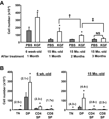
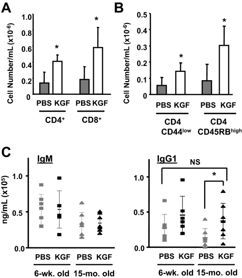
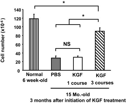
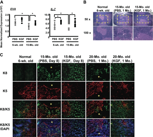
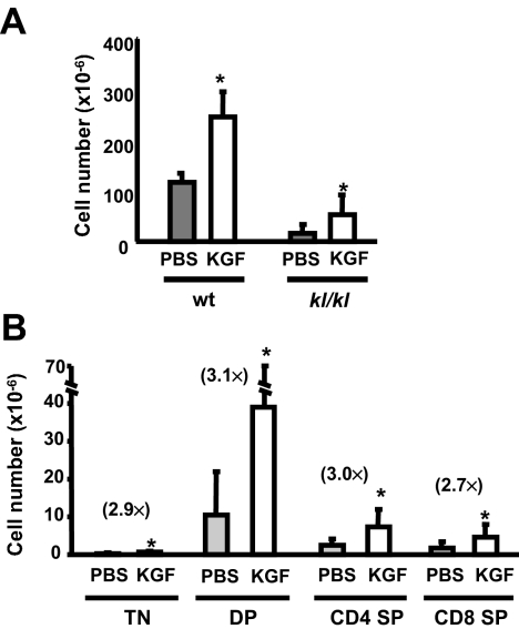
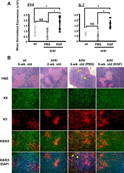
Age-related thymopoietic insufficiency has been proposed to be related to either defects in lymphohematopoietic progenitors or the thymic microenvironment. In this study, we examined whether keratinocyte growth factor (KGF), an epithelial cell-specific growth factor, could increase thymopoietic capacity in aged mice by restoration of the function of thymic epithelial cells (TECs). The thymic cellularity in KGF-treated aged mice increased about 4-fold compared to placebo-treated mice, resulting in an equivalent thymic cellularity to young mice. Enhanced thymopoiesis was maintained for about 2 months after a single course of KGF, and sustained improvement was achieved by administration of monthly courses of KGF. With the enhanced thymopoiesis after KGF treatment, the number of naive CD4 T cells in the periphery and T-cell-dependent antibody production improved in aged mice. KGF induced increased numbers of TECs and intrathymic interleukin-7 (IL-7) production and reorganization of cortical and medullary architecture. Furthermore, KGF enhanced thymopoiesis and normalized TEC organization in klotho (kl/kl) mice, a model of premature degeneration and aging, which displays thymopoietic defects. The result suggests that TEC damage is pathophysiologically important in thymic aging, and KGF therapy may be clinically useful in improving thymopoiesis and immune function in the elderly.
Figures
Similar articles
-
Protection from thymic epithelial cell injury by keratinocyte growth factor: a new approach to improve thymic and peripheral T-cell reconstitution after bone marrow transplantation.Blood. 2002 Jun 15;99(12):4592-600. doi: 10.1182/blood.v99.12.4592. Blood. 2002. PMID: 12036893
-
Keratinocyte growth factor preserves normal thymopoiesis and thymic microenvironment during experimental graft-versus-host disease.Blood. 2002 Jul 15;100(2):682-91. doi: 10.1182/blood.v100.2.682. Blood. 2002. PMID: 12091365
-
Short-term inhibition of p53 combined with keratinocyte growth factor improves thymic epithelial cell recovery and enhances T-cell reconstitution after murine bone marrow transplantation.Blood. 2010 Feb 4;115(5):1088-97. doi: 10.1182/blood-2009-05-223198. Epub 2009 Dec 4. Blood. 2010. PMID: 19965631 Free PMC article.
-
Thymic involution and immune reconstitution.Trends Immunol. 2009 Jul;30(7):366-73. doi: 10.1016/j.it.2009.04.003. Epub 2009 Jun 18. Trends Immunol. 2009. PMID: 19540807 Free PMC article. Review.
-
Thymic Epithelial Cells Contribute to Thymopoiesis and T Cell Development.Front Immunol. 2020 Jan 31;10:3099. doi: 10.3389/fimmu.2019.03099. eCollection 2019. Front Immunol. 2020. PMID: 32082299 Free PMC article. Review.
Cited by
-
A GMCSF and IL7 fusion cytokine leads to functional thymic-dependent T-cell regeneration in age-associated immune deficiency.Clin Transl Immunology. 2015 May 8;4(5):e37. doi: 10.1038/cti.2015.8. eCollection 2015 May. Clin Transl Immunology. 2015. PMID: 26131365 Free PMC article.
-
Administration of recombinant FOXN1 protein attenuates Alzheimer's pathology in mice.Brain Behav Immun. 2023 Oct;113:341-352. doi: 10.1016/j.bbi.2023.07.027. Epub 2023 Aug 2. Brain Behav Immun. 2023. PMID: 37541395 Free PMC article.
-
The effect of age on thymic function.Front Immunol. 2013 Oct 7;4:316. doi: 10.3389/fimmu.2013.00316. Front Immunol. 2013. PMID: 24109481 Free PMC article. Review.
-
Declining expression of a single epithelial cell-autonomous gene accelerates age-related thymic involution.Aging Cell. 2010 Jun;9(3):347-57. doi: 10.1111/j.1474-9726.2010.00559.x. Epub 2010 Mar 12. Aging Cell. 2010. PMID: 20156205 Free PMC article.
-
Nonhuman primate models of human immunology.Antioxid Redox Signal. 2011 Jan 15;14(2):261-73. doi: 10.1089/ars.2010.3241. Epub 2010 Aug 30. Antioxid Redox Signal. 2011. PMID: 20524846 Free PMC article. Review.
References
-
- Jamieson BD, Douek DC, Killian S, et al. Generation of functional thymocytes in the human adult. Immunity. 1999;10:569–575. - PubMed
-
- Mackall CL, Fleisher TA, Brown MR, et al. Age, thymopoiesis, and CD4+ T lymphocyte regeneration after intensive chemotherapy. New Engl J Med. 1995;332:143–149. - PubMed
-
- Douek DC, McFarland RD, Keiser PH, et al. Changes in thymic function with age and during the treatment of HIV infection. Nature. 1998;396:690–695. - PubMed
-
- Small TN, Papadopoulos EB, Boulad F, et al. Comparison of immune reconstitution after unrelated and related T-cell-depleted bone marrow transplantation: effect of patient age and donor leukocyte infusions. Blood. 1999;93:467–480. - PubMed
Publication types
MeSH terms
Substances
Grants and funding
- R01 HL073794/HL/NHLBI NIH HHS/United States
- AG25326/AG/NIA NIH HHS/United States
- A1057477/PHS HHS/United States
- AG19712/AG/NIA NIH HHS/United States
- HL073794/HL/NHLBI NIH HHS/United States
- R01 HL070005/HL/NHLBI NIH HHS/United States
- HL70005/HL/NHLBI NIH HHS/United States
- HL54729/HL/NHLBI NIH HHS/United States
- R01 AG019712/AG/NIA NIH HHS/United States
- R01 AG025326/AG/NIA NIH HHS/United States
- P01 HL073104/HL/NHLBI NIH HHS/United States
- HL73104/HL/NHLBI NIH HHS/United States
- R01 HL054729/HL/NHLBI NIH HHS/United States
LinkOut - more resources
Full Text Sources
Other Literature Sources
Medical
Molecular Biology Databases
Research Materials
