. 2006 Dec 13;25(24):5726-34.
doi: 10.1038/sj.emboj.7601453. Epub 2006 Nov 30.

Repression of ADH1 and ADH3 during zinc deficiency by Zap1-induced intergenic RNA transcripts

Affiliations

Repression of ADH1 and ADH3 during zinc deficiency by Zap1-induced intergenic RNA transcripts

Amanda J Bird et al. EMBO J. .

Abstract

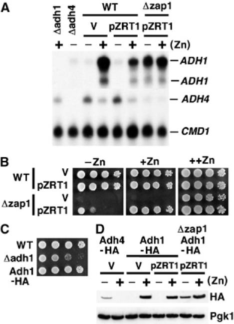
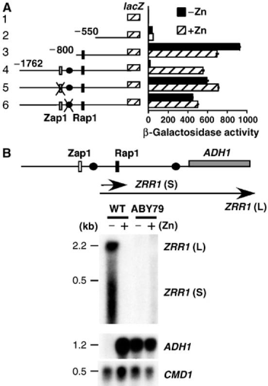
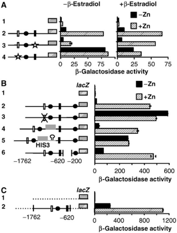
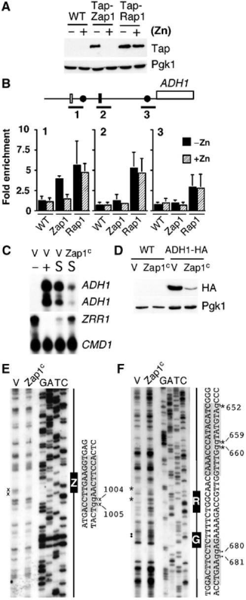
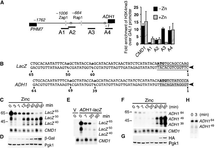
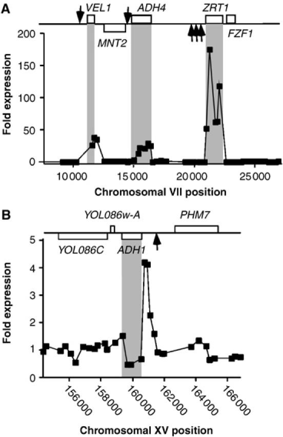
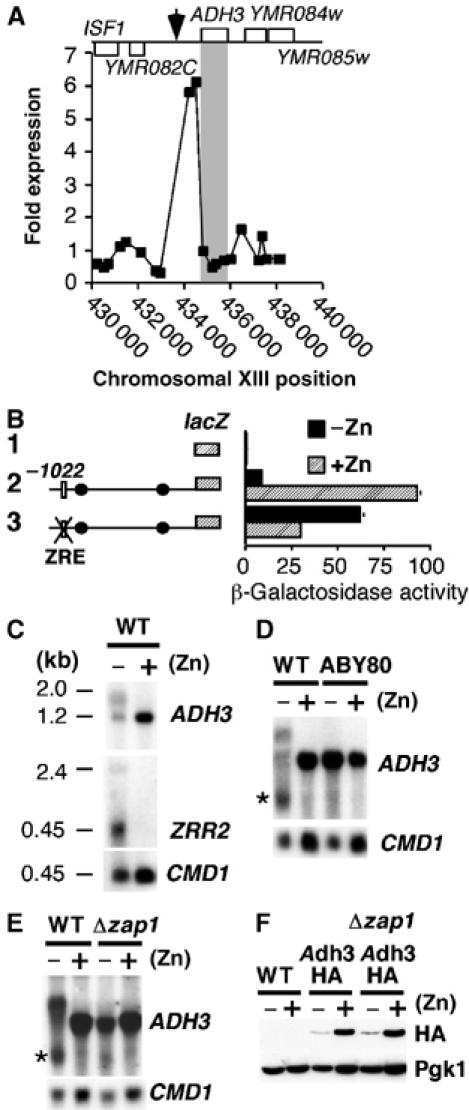
The transcriptional activator Zap1 induces target gene expression in response to zinc deficiency. We demonstrate that during zinc starvation, Zap1 is required for the repression of ADH1 expression. ADH1 encodes the major zinc-dependent alcohol dehydrogenase that is utilized during fermentation. During zinc starvation, Zap1 binds upstream of the activator Rap1 and induces an intergenic RNA transcript, ZRR1. ZRR1 expression leads to the transient displacement of Rap1 from the ADH1 promoter resulting in ADH1 repression. Using a microarray-based approach, we screened for additional genes repressed by Zap1 intergenic transcripts. We found that ADH3, the major mitochondrial alcohol dehydrogenase, is regulated in a manner similar to ADH1. Thus, during zinc deficiency, Zap1 mediates the repression of two of the most abundant zinc-requiring enzymes.


Figures

Figure 1
Figure 1
Differential regulation of ADH1 and ADH4 expression in response to zinc. The wild-type strain BY4741 and zap1Δmutant strain were transformed with pZRT1 or the empty vector (V). Total RNA was extracted following growth to exponential phase in LZM media supplemented with either 5 μM zinc [−Zn] or 3000 μM zinc [+Zn] and the levels of ADH4 and ADH1 analyzed using S1 nuclease analysis (A). RNA from adh4Δ and adh1Δ mutant strains was used to confirm the specificity of the probes, whereas calmodulin (CMD1) was used as a loading control. The strains BY4741 and zap1Δ containing the indicated plasmids were grown overnight in synthetic deplete medium lacking uracil (SD –URA). Five microliters of a cell suspension (OD600 1.0) and three 10-fold serial dilutions were plated onto SD (−URA) medium [++Zn] or SD medium (−uracil) containing 1 mM EDTA supplemented with 50 μM zinc [+Zn] or 3 μM zinc [−Zn]. Plates were grown for 3 days at 30°C (B). The strains BY4741 (WT), adh1Δ and ABY69 (Adh1-HA) were grown in synthetic complete (SC) medium overnight before serial dilutions were made onto SC medium, as described in panel B (C). The strains BY4741 (WT), ABY64 (Adh4-HA), ABY69 (Adh1-HA) and ABY77 (Δzap1, Adh1-HA) containing the indicated plasmids were grown as described in panel A. Crude protein extracts were made and analyzed by immunoblotting. Phosphoglycerate kinase 1 (Pgk1) was used as a loading control (D).
Figure 2
Figure 2
ADH1 repression requires the Zap1 dependent RNA transcript ZRR1. The indicated reporter constructs were transformed into in the wild-type strain BY4741. Transformants were grown to exponential phase in LZM supplemented with either 5 μM zinc [−Zn] or 3000 μM zinc [+Zn] and β-galactosidase activity measured in triplicate by standard procedures (A). The ZRE (white rectangle), Rap1 UAS (black rectangle) and upstream TATA box (black oval) are shown. Mutation of the ZRE element (ACCTTGAAGGT to TGGTTGAAGGT) or TATA box sequence (TATAAA to CCTAGG) is indicated by a cross. All numbers are relative to the first base of the initiation codon of lacZ, which is designated as +1. Northern analysis of ADH1, ZRR1 and loading control CMD1. The wild-type strain BY4741 and strain ABY79 in which the ADH1-ZRE (−1006 to −995 bp) is replace by the K. lactis URA3 ORF, were grown as described in panel A (B). The northern probe for ADH1 was a 54 bp oligonucleotide that hybridized to −27 to +27 bp relative to the ATG translational codon.
Figure 3
Figure 3
Characterization of the promoter elements required for ADH1 repression. The strain YM4271 (gal4Δ) was transformed with the indicated constructs. Transformed cells were grown as described in Figure 2A in the presence or absence of 10–5 M β-estradiol. β-Galactosidase activity measured in triplicate by standard procedures (A). The indicated reporter constructs were transformed into the wild-type strain BY4741 and cells grown as described in Figure 2A. β-Galactosidase activity measured in triplicate by standard procedures. The HIS3 ORF (gray box), HIS3 terminator (hairpin loop), and CYC1 promoter region (dashed line) and Gal4 UAS (star) are shown. All other symbols are described in Figure 2A (B, C).
Figure 4
Figure 4
Expression of ZRR1 leads to the transient dissociation of Rap1 from the ADH1 promoter. The strains BY4741, ABY61 (TAP-Zap1) and TAP-Rap1 were grown to exponential phase in LZM supplemented with either 3 μM zinc [−Zn] or 3000 μM zinc [+Zn]. (A) Crude protein extracts were generated and analyzed by immunoblotting. Blots were incubated with peroxidase–anti peroxidase (for TAP) or Pgk1 for the loading control. (B) Cells grown under the conditions in panel A were crosslinked for ChIP analysis. PCR was performed with primer pairs that amplified the following sequences relative to the first base of the initiation codon of ADH1, which is designated as +1 (bar 1 −1163 to −778 bp, bar 2 −837 to −486 bp and bar 3 −290 to +58 bp). Amplified sequences were normalized against input DNA and binding at the HIS3 ORF (negative control). Data represents an average of three independent experiments. The extension of Rap1 binding in bar 1 and bar 3 sequences likely arises from incomplete sonication. (C) The strain BY4741 containing the empty vector pRS-VP16 (V) or VP16-ZAP1 (Zap1c) was grown to exponential phase in LZM supplemented with 5 μM zinc (−), 3000 μM zinc (+) or in Zn-replete SD (−URA) (S). Total RNA was extracted and the levels of ADH1, ZRR1 and CMD1 mRNAs were analyzed by S1 nuclease analysis. (D) The strains BY4741 and ABY69 (ADH1-HA) containing pRS-VP16 (V) or Zap1c were grown in SD (−URA). Crude protein extracts were made and analyzed by immunoblotting. (E, F) Exponential cultures of BY4741 containing the above plasmids were grown to exponential phase in SD (−URA) media and analyzed by in vivo DMS footprinting. The positions of the ZRE (Z), Rap1 (R) and Gcr1 (G) binding sites have been indicated schematically and by gray shading in the DNA sequence. Protected G residues have been indicated by × (Zap1), * (Rap1), • (Gcr1) and by lowercase letters in the DNA sequence. Data is a representative of 3 independent experiments.
Figure 5
Figure 5
Effects of ZRR1 on ADH1 transcriptional initiation. The wild-type strain BY4741 was grown under conditions described in Figure 4A. ChIP was performed with anti-histone H3K4me3 antibodies and real time quantitative PCR performed with PCR primers that amplified the following sequences relative to the ADH1 ATG codon; A1 −1136 to −949 bp, A2 −974 to −820 bp, A3 −479 to −345 bp, A4 −94 to +84 bp. The CMD1 control primers amplified sequences −227 to +61 bp relative to the CMD1 ATG codon. Values are an average of three quantifications (A). DNA sequence of the S1 oligonucleotides used in panels C–F. Arrowheads indicate the 5′ ends of the oligonucleotides. Shown are the complementary DNA strands (the positions of the previously mapped –27 and –37 ADH1 transcriptional start site (bold lowercase letters) (Bennetzen and Hall, 1982), the translational ATG (bold uppercase letters), the ADH1 ORF (gray shading), the TATA boxes (open ovals) and lacZ ORF (underlined sequence). Numbers indicate the base-pair position in the oligonucleotide (B). Strain BY4741 containing the integrated ADH1-lacZ fusion (Figure 2A construct 4) was grown to exponential phase in LZM media supplemented with 5 μM Zn2+. Cells were harvested, washed 3 times in PBS and then resuspended in SD (−URA) supplemented with 100 μM Zn2+. Aliquots were removed at the indicated time points for S1 nuclease analysis (C) or immunoblot analysis (D). The strain ABY79 (ΔADH1ZRE) containing pYep353 or pYepADH1 (multi-copy version of Figure 2 construct 4) was grown as described in panel C. Aliquots were removed at the indicated times for S1 nuclease analysis (E). Strain ABY69 (ADH1-HA) was grown as described in panel C. Cells were removed for S1 nuclease analysis (F) or immunoblot analysis (G). A longer exposure of lanes 1 and 2 of panel F was performed (H).
Figure 6
Figure 6
Identification of additional Zap1-dependent intergenic RNA transcripts. Shown are data for the ZRT1 (A) and ADH1 (B) chromosomal regions. The strain zap1Δ containing either pRS-VP16 (vector) or pZap1c was grown to exponential phase in SD (−uracil) media before cells were harvested. RNA was extracted and hybridized to yeast intergenic microarrays as described in Supplementary Figure 2. The differential expression between the vector and pZap1c labeled samples was plotted against chromosomal map position. The positions of ORFs have been indicated where upper boxes represent the Watson strand ORFs and lower boxes represent the Crick strand. Arrows indicate the positions of ZREs. Gray panels highlight the positions of Zap1 regulated ORFs.
Figure 7
Figure 7
ADH3 gene expression is regulated by the Zap1-dependent ZRR2 transcript. (A) Expression differences in the strain zap1Δ containing either pRS-VP16 (vector) or pZap1c. See legend of Figure 4C and D for details. (B) The indicated reporter constructs were transformed into the wild-type strain BY4741 and cells grown to exponential phase in LZM supplemented with either 3 μM zinc [−Zn] or 3000 μM zinc [+Zn] and β-galactosidase activity measured in triplicate by standard procedures. (C–E) Northern analysis of ADH3, ZRR2 and loading control CMD1. Wild-type strain BY4741, strain ABY80 in which the ADH3-ZRE is replaced by the K. lactis URA3 ORF (−755 to –745 bp relative to the ATG) or the zap1Δ mutant strain containing pZRT1 (Δzap1) was grown under the conditions described in panel B. The northern probe for ADH3 was a 60 bp oligonucleotide that hybridized to +6 to +65 bp relative to the ATG translational codon. (F) The strains BY4741 (WT), ABY71 (Adh3-HA) and ABY74 (Δzap1 Adh3-HA) were grown in LZM media supplemented with either 3 μM zinc [−Zn] or 3000 μM zinc [+Zn]. Crude protein extracts were made and analyzed by immunoblotting. Phosphoglycerate kinase 1 (Pgk1) was used as a loading control.

References

    1. Bennetzen JL, Hall BD (1982) The primary structure of the Saccharomyces cerevisiae gene for alcohol dehydrogenase. J Biol Chem 257: 3018–3025 - PubMed
    1. Bird AJ, Blankman E, Stillman DJ, Eide DJ, Winge DR (2004) The Zap1 transcriptional activator also acts as a repressor by binding downstream of the TATA box in ZRT2. EMBO J 23: 1123–1132 - PMC - PubMed
    1. Bird AJ, McCall K, Kramer M, Blankman E, Winge DR, Eide DJ (2003) Zinc fingers can act as Zn2+ sensors to regulate transcriptional activation domain function. EMBO J 22: 5137–5146 - PMC - PubMed
    1. Bird AJ, Swierczek S, Qiao W, Eide DJ, Winge DR (2006) Zinc metalloregulation of the zinc finger pair domain. J Biol Chem 281: 25326–25335 - PubMed
    1. Bird AJ, Zhao H, Luo H, Jensen LT, Srinivasan C, Evans-Galea M, Winge DR, Eide DJ (2000) A dual role for zinc fingers in both DNA binding and zinc sensing by the Zap1 transcriptional activator. EMBO J 19: 3704–3713 - PMC - PubMed

Publication types

MeSH terms