. 2006 Dec 13;25(24):5961-9.
doi: 10.1038/sj.emboj.7601452. Epub 2006 Nov 30.

Structure of the origin-binding domain of simian virus 40 large T antigen bound to DNA

Affiliations

Structure of the origin-binding domain of simian virus 40 large T antigen bound to DNA

Elena Bochkareva et al. EMBO J. .

Abstract

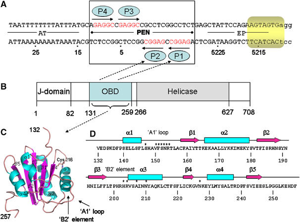
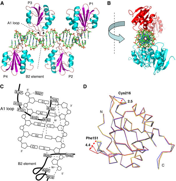
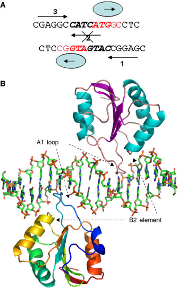
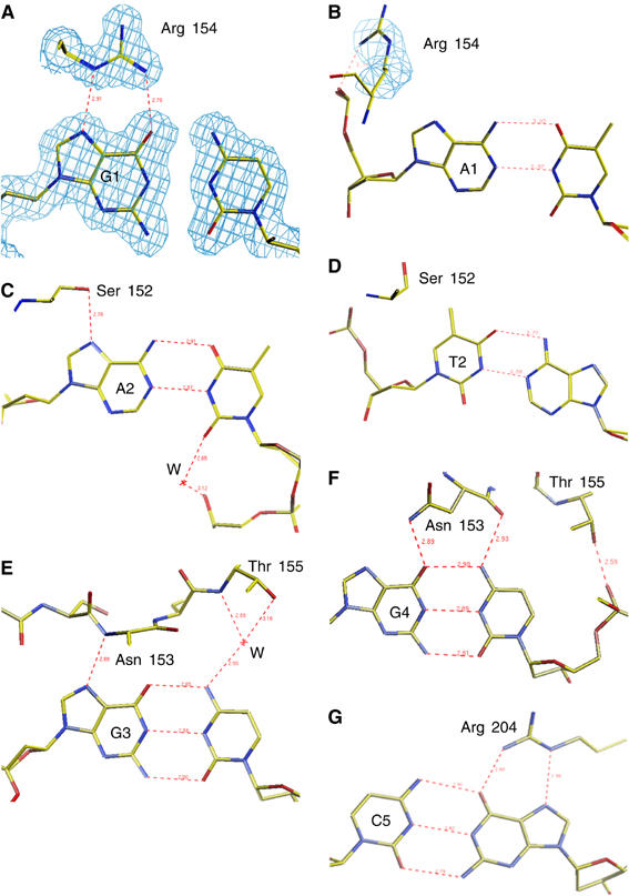
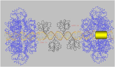
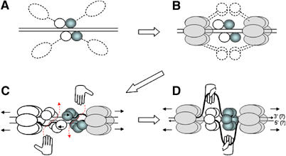
The large T antigen (T-ag) protein binds to and activates DNA replication from the origin of DNA replication (ori) in simian virus 40 (SV40). Here, we determined the crystal structures of the T-ag origin-binding domain (OBD) in apo form, and bound to either a 17 bp palindrome (sites 1 and 3) or a 23 bp ori DNA palindrome comprising all four GAGGC binding sites for OBD. The T-ag OBDs were shown to interact with the DNA through a loop comprising Ser147-Thr155 (A1 loop), a combination of a DNA-binding helix and loop (His203-Asn210), and Asn227. The A1 loop traveled back-and-forth along the major groove and accounted for most of the sequence-determining contacts with the DNA. Unexpectedly, in both T-ag-DNA structures, the T-ag OBDs bound DNA independently and did not make direct protein-protein contacts. The T-ag OBD was also captured bound to a non-consensus site ATGGC even in the presence of its canonical site GAGGC. Our observations taken together with the known biochemical and structural features of the T-ag-origin interaction suggest a model for origin unwinding.


Figures

Figure 1
Figure 1
Structural and functional elements of SV40 DNA replication system. (A) Sequences present in the 64-bp core origin of SV40 replication. Locations of the AT-rich region, the pentanucleotide box (PEN) and EP sites, numbered as in the genome of reference strain 776. Arrows depict the four GAGGC pentanucleotides—the binding sites for T-ag OBDs (blue ellipses). Eight nucleotides within the EP that melt upon the T-ag double hexamer assembly are highlighted in yellow. The 23 bp PEN element structure reported in this work is boxed. (B) Functional domains of SV40 large T antigen (T-ag). ‘J Domain', region not required for in vitro DNA replication; ‘OBD' origin DNA-binding domain, minimal region required for binding to SV40 Ori DNA; ‘Helicase', minimal region required for the helicase activity; the numbers given are the amino-acid residues. (C) T-ag OBD structure (apo form) ribbon diagram showing the domain containing residues 134–259 (in figure it is 132–257). α-Helices are in cyan and β-strands in magenta. Position of Cys 216 and DNA-binding components ‘A1 loop' and ‘B2 element' are indicated. (D) Secondary structure elements of T-ag OBD. The β-strands are indicated by arrows and the α-helices by boxes. Amino acids contributing to DNA binding are labeled with ‘*'.
Figure 2
Figure 2
Structure of four T-ag OBDs bound to the 23 bp PEN box of ori DNA (PEN-4). (A) Four copies of OBD are represented as ribbon models and colored as in Figure 1C. DNA is shown as a stick model. (B) Same structure, PEN-4, viewed along the DNA axis. OBDs bound to sites 1 and 3 are colored in cyan and those bound to sites 2 and 4 are in red. (C) A schematic diagram showing the contacts of T-ag OBD with the GAGGC pentanucleotide. The bases are numbered 1–5 starting from the 5′ end of the recognition element. The amino acids are boxed and the trace of polypeptide backbone is indicated with a thick dark line. The broken lines indicate hydrogen bonds. ‘W' for water molecules. (D) Conformational changes in T-ag OBD induced by DNA binding. A superposition of free and bound OBD was generated as discussed in the text. Shown is the Cα trace of the free (blue and yellow for the P21 and P212121 crystal forms, respectively) and bound (red) domain. Important amino acids are labeled. Maximal shift of the loops are indicated with dashed lines, and the size of the shift is indicated in Å (large numbers).
Figure 3
Figure 3
Non-sequence-specific binding of T-ag OBD to PEN-2. (A) DNA used for cocrystallization of the complex. Arrows depict two GAGGC pentanucleotides, sites 1 and 3. Mutated site 2 is crossed out. Non-canonical binding sites bound by T-ag OBD in the structure are shown in red. (B) T-ag OBD–PEN-2 complex structure. Two copies of OBD are represented as ribbon models. One monomer is colored according to the secondary structure; α-helices in cyan and β-strands in purple. The other monomer is rainbow colored from blue (N-terminus) to red (C-terminus).
Figure 4
Figure 4
Structural details of T-ag interaction with DNA bases in the PEN-2 and PEN-4 structures. (A) Sequence-specific interaction of Arg 154 with G1 (the first G in the GAGGC pentamer) in the PEN-4 structure. A representative electron density map (shown in blue) is superimposed on the model. (B) Nonspecific interaction of Arg 154 with A1 (A in position of G1) in the PEN-2 structure. A representative electron density is superimposed on Arg 154. (C) Sequence-specific interaction of Ser 152 with A2. (D) Nonspecific interaction of Ser 152 with the T2 in the PEN-2 structure. Sequence-specific interactions, which involve (E) the G3 and Asn 153, (F) the G4 and Asn 153, and (G) the G (complementary to C5) with Arg 204. The protein and DNA are shown as stick models and colored by atom type; yellow for carbon, blue for nitrogen, red for oxygen, and purple for phosphorus. A representative electron density as captured from 2FoFc map is shown with contours drawn at the 1.25σ level. Hydrogen bonds are indicated with red dashed lines, and the length of the bonds is indicated in Å.
Figure 5
Figure 5
Molecular model of an initial step in the SV40 DNA replication. The PEN-4 structure is shown in black, two hexameric helicase domains are in blue (PDB Id: 1N25), modeled DNA is shown as a stick model and colored per atom type (carbon in yellow, oxygen in red, nitrogen in blue, and phosphorus in purple). Position of the initially melted 8 nt fragment of EP with respect to the PEN box is highlighted with a yellow rectangle. Relative positions of the C-terminus in the OBD (aa 253) and N-terminus in the helicase domain (aa 266) are indicated. See text for more detail.
Figure 6
Figure 6
Model for origin melting and DNA unwinding by T-ag. (A) Four OBDs bind the origin and bring monomeric domains of helicase in proximity to the DNA. (B) Helicase domains are assembled in two hexamers encircling DNA; this brings an additional eight OBDs in proximity to the PEN box. (C) Using a nonspecific DNA-binding mode, the eight additional OBDs bind side by side with the four initial OBDs forming a spiral double hexamer. The moving helicase distorts and melts a fragment of EP, making it accessible for RPA (hands). RPA moves a melted strand of DNA out through the open gate (red dashed arrow). (D) The moving helicase pulls the most distant OBD (via the short linker comprising aa 253–266) and induces switching of an open (spiral) OBD hexamer to closed hexamer. OBDs are represented as circles. OBDs that mediate assembly of the right-hand hexamer are colored in blue and those for the left-hand hexamer are white. Monomers of the helicase domain are represented as ellipses and shadowed in gray. The direction of helicase movement is indicated by detached arrows in front of the helicase hexamers. A putative polarity of the DNA strands is shown by small arrows at the DNA ends.

References

    1. Arthur AK, Hoss A, Fanning E (1988) Expression of simian virus 40T antigen in Escherichia coli: localization of T-antigen origin DNA-binding domain to within 129 amino acids. J Virol 62: 1999–2006 - PMC - PubMed
    1. Bochkarev A, Barwell JA, Pfuetzner RA, Bochkareva E, Frappier L, Edwards AM (1996) Crystal structure of the DNA-binding domain of the Epstein–Barr virus origin-binding protein, EBNA1, bound to DNA. Cell 84: 791–800 - PubMed
    1. Bochkarev A, Barwell JA, Pfuetzner RA, Furey W Jr, Edwards AM, Frappier L (1995) Crystal structure of the DNA-binding domain of the Epstein–Barr virus origin-binding protein EBNA 1. Cell 83: 39–46 - PubMed
    1. Borowiec JA, Hurwitz J (1988) Localized melting and structural changes in the SV40 origin of replication induced by T-antigen. EMBO J 7: 3149–3158 - PMC - PubMed
    1. Bullock PA (1997) The initiation of simian virus 40 DNA replication in vitro. Crit Rev Biochem Mol Biol 32: 503–568 - PubMed

Publication types