

Normal and abnormal transformation of the spiral arteries during pregnancy
- PMID: 17140293
- PMCID: PMC7062302
- DOI: 10.1515/JPM.2006.089
Normal and abnormal transformation of the spiral arteries during pregnancy
Abstract
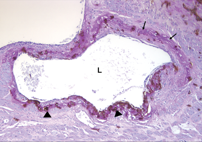
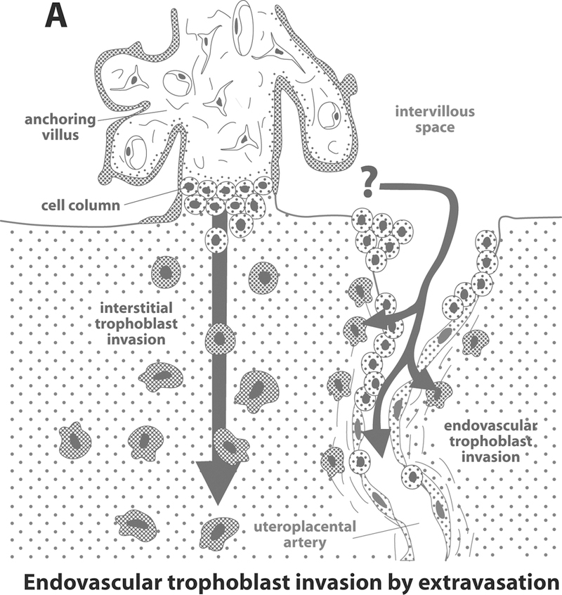
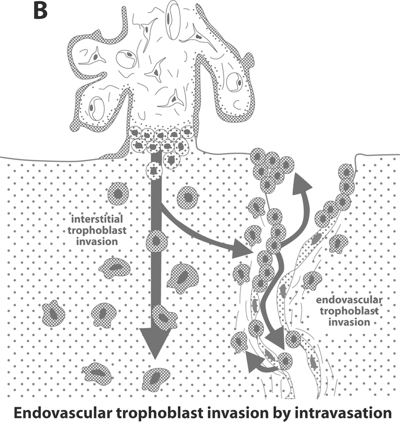
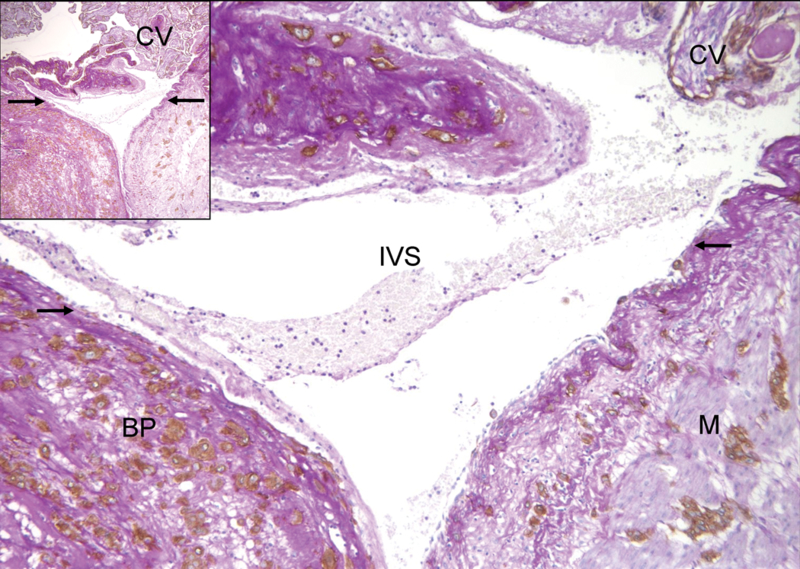
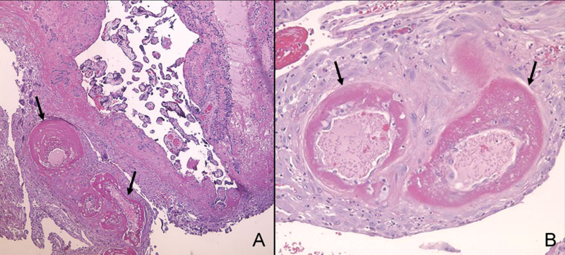
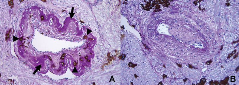
This article reviews the anatomy and physiology of the uterine circulation, with emphasis on the remodeling of spiral arteries during normal pregnancy, and the timing and anatomical pathways of trophoblast invasion of the spiral arteries. We review the definitions of the placental bed and basal plate of the placenta, their relevance to the study of the physiologic transformation of the spiral arteries, as well as the methods to obtain and examine placental bed biopsy specimens. We also examine the role of the extravillous trophoblast in normal and abnormal pregnancies, and the criteria used to diagnose failure of physiologic transformation of the spiral arteries. Finally, we comment on the use of uterine artery Doppler velocimetry as a surrogate marker of chronic uteroplacental ischemia.
Figures
References
-
- Aardema MW, Oosterhof H, Timmer A, van R, I, Aarnoudse JG: Uterine artery Doppler flow and uteroplacental vascular pathology in normal pregnancies and pregnancies complicated by pre-eclampsia and small for gestational age fetuses. Placenta 22 (2001) 405. - PubMed
-
- Abramowsky CR, Vegas ME, Swinehart G, Gyves MT: Decidual vasculopathy of the placenta in lupus erythematosus. N Engl J Med 303 (1980) 668. - PubMed
-
- Albaiges G, Missfelder-Lobos H, Lees C, Parra M, Nicolaides KH: One-stage screening for pregnancy complications by color Doppler assessment of the uterine arteries at 23 weeks’ gestation. Obstet Gynecol 96 (2000) 559. - PubMed
-
- Alberts B, Bray D, Lewis J, Raff M, Roberts K, Watson JD: Cell junctions, cell adhesion, and the extracellular matrix. In: Molecular biology of the cell. 1994
-
- Arduini D, Rizzo G, Romanini C, Mancuso S: Utero-placental blood flow velocity waveforms as predictors of pregnancy-induced hypertension. Eur J Obstet Gynecol Reprod Biol 26 (1987) 335. - PubMed
Publication types
MeSH terms
Grants and funding
LinkOut - more resources
Full Text Sources
Medical