. 2007 Mar;120(3):354-61.
doi: 10.1111/j.1365-2567.2006.02510.x. Epub 2006 Nov 28.

Functional characterization of a T-cell receptor BV6+ T-cell clone derived from a leprosy lesion

Affiliations

Functional characterization of a T-cell receptor BV6+ T-cell clone derived from a leprosy lesion

Shereen Sabet et al. Immunology. 2007 Mar.

Abstract

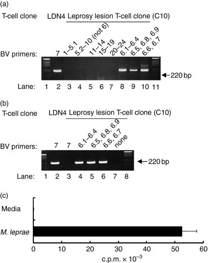
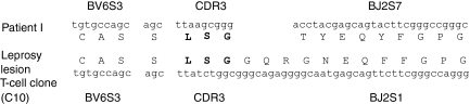
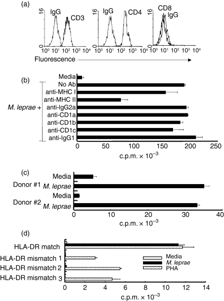
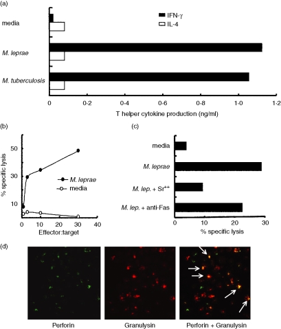
Human infection with Mycobacterium leprae, an intracellular bacterium, presents as a clinical and immunological spectrum; thus leprosy provides an opportunity to investigate mechanisms of T-cell responsiveness to a microbial pathogen. Analysis of the T-cell receptor (TCR) repertoire in leprosy lesions revealed that TCR BV6(+) T cells containing a conserved CDR3 motif are over-represented in lesions from patients with the localized form of the disease. Here, we derived a T-cell clone from a leprosy lesion that expressed TCR BV6 and the conserved CDR3 sequence L-S-G. This T-cell clone produced a T helper type 1 cytokine pattern, directly lysed M. leprae-pulsed antigen-presenting cells by the granule exocytosis pathway, and expressed the antimicrobial protein granulysin. BV6(+) T cells may therefore functionally contribute to the cell-mediated immune response against M. leprae.


Figures

Figure 1
Figure 1
PCR analysis of a BV6+ T-cell clone. (a) Pools of BV primers at equal concentrations were used to assess the BV chain gene usage of a leprosy-lesion-derived T-cell clone (C10); lanes 1 and 11 contain the 1-kb ladder marker; lane 2 contains BV7-amplified cDNA from a BV7-expressing T-cell clone, LDN4; pools of primers are as follows, lane 3, BVs 1, 2, 3, 4, 5.1; lane 4, BVs 5.23, 7, 8, 9, 10; lane 5, BVs 11, 12, 13.1, 13.2, 14; lane 6, BVs 15, 16, 17, 18, 19; lane 7, BVs 20, 21, 22, 23, 24; lane 8, BV 6.1/2/3/4; lane 9, BV 6.5/8/9; lane 10, BV 6.6/7. (b) Lane 2, BV7+ T-cell clone; lane 3, leprosy lesion clone (C10) plus BV7 primer (as a negative control); lanes 4, 5 and 6, leprosy lesion clone (C10) plus BV 6.1/2/3/4, 6.5/8/9, and 6.6/7 primers; lane 7, leprosy lesion clone with no BV primers (negative control lanes). (c) Proliferative response of a leprosy lesion T-cell clone to M. leprae antigen. The BV6+ T-cell clone was stimulated with M. leprae bacterial lysates in a [3H]thymidine incorporation assay. The data represent one of more than 30 experiments. Values are expressed as the mean ± SEM of triplicate cultures.
Figure 2
Figure 2
Sequence analysis of the BV6+ T-cell clone and comparison to a previously identified T-Lep patient BV6+ TCR. The T-cell clone shares the same BV chain as a patient TCR from a previous study. The T-cell clone also expresses the exact CDR3 ‘L-S-G’ motif, but uses a different BJ gene.
Figure 3
Figure 3
Co-receptor expression and MHC restriction of BV6+ T-cell clone. (a) Phenotypic analysis of the BV6+ T-cell clone was performed by flow cytometry. Isotype control and specific antibodies are labelled on the histograms. (b) APCs were cultured with neutralizing antibodies to MHC and MHC-like antigen-presenting molecules (60 min) before T cells were added. Data shown are representative of three experiments. Values are expressed as the mean ± SEM of triplicate cultures. (c) APCs were from two HLA-DR-matched individuals. Data shown are representative of three experiments. (d) T-cell clone does not respond to antigen presented by HLA-DR mismatched APCs. Values are expressed as the mean ± SEM of triplicate cultures. Data shown are representative of multiple experiments.
Figure 4
Figure 4
T-cell effector functions of the BV6+ T-cell clone. (a) Cytokines (IFN-γ and IL-4) were measured from supernatants of T cells stimulated with antigen (M. leprae or M. tuberculosis) or media. The data are presented as the mean of triplicate wells and are representative of three experiments. (b) Cytolytic analysis of the BV6+ T-cell clone. Leprosy lesion T-cell clones can affect cytolytic activity against M. leprae-pulsed APCs as measured in a 51Cr-release assay. The data are representative of four separate experiments. (c) T cells were incubated with strontium (18 hr) to degranulate cytolytic mediators or with anti-Fas antibody to prevent Fas–FasL interactions. Monocyte-derived dendritic cells were then incubated with T cells. Cytolytic activity of the T-cell clones measured by a 51Cr-release assay. The data are representative of three experiments. (d) Two-colour immunofluorescent cell labelling of the BV6+ T-cell clone reveals that both granulysin and perforin co-localize in individual cells (white arrows).

References

    1. Leprosy. Global situation. Wkly Epidemiol Rec. 2002;77:1–8. - PubMed
    1. Salgame P, Abrams JS, Clayberger C, Goldstein H, Convit J, Modlin RL, Bloom BR. Differing lymphokine profiles of functional subsets of human CD4 and CD8 T cell clones. Science. 1991;254:279–82. - PubMed
    1. Yamamura M, Uyemura K, Deans RJ, Weinberg K, Rea TH, Bloom BR, Modlin RL. Defining protective responses to pathogens: cytokine profiles in leprosy lesions. Science. 1991;254:277–9. - PubMed
    1. Modlin RL, Melancon-Kaplan J, Young SMM, Pirmez C, Kino H, Convit J, Rea TH, Bloom BR. Learning from lesions: patterns of tissue inflammation in leprosy. Proc Natl Acad Sci USA. 1988;85:1213–17. - PMC - PubMed
    1. Wang X, Golkar L, Uyemura K, et al. T cells bearing Vβ6 T-cell receptors in the cell-mediated immune response to Mycobacterium leprae. J Immunol. 1993;151:7105–16. - PubMed

Publication types

MeSH terms

Substances